Conexiant
Login
  • The Analytical Scientist
  • The Cannabis Scientist
  • The Medicine Maker
  • The Ophthalmologist
  • The Pathologist
  • The Traditional Scientist
The Analytical Scientist
  • Explore

    Explore

    • Latest
    • News & Research
    • Trends & Challenges
    • Keynote Interviews
    • Opinion & Personal Narratives
    • Product Profiles
    • App Notes

    Featured Topics

    • Mass Spectrometry
    • Chromatography
    • Spectroscopy

    Issues

    • Latest Issue
    • Archive
  • Topics

    Techniques & Tools

    • Mass Spectrometry
    • Chromatography
    • Spectroscopy
    • Microscopy
    • Sensors
    • Data and AI

    • View All Topics

    Applications & Fields

    • Clinical
    • Environmental
    • Food, Beverage & Agriculture
    • Pharma and Biopharma
    • Omics
    • Forensics
  • People & Profiles

    People & Profiles

    • Power List
    • Voices in the Community
    • Sitting Down With
    • Authors & Contributors
  • Business & Education

    Business & Education

    • Innovation
    • Business & Entrepreneurship
    • Career Pathways
  • Events
    • Live Events
    • Webinars
  • Multimedia
    • Video
Subscribe
Subscribe

False

The Analytical Scientist / App Notes / 2016 / Confocal Raman Imaging in Life Science - Living Cells, Bacteria & Tissues

Confocal Raman Imaging in Life Science - Living Cells, Bacteria & Tissues

04/22/2016

Share

Featured Image

From measurements in liquids, to solid samples and soft tissues in life science research various samples are analysed on a regular basis. Through their convenient handling and versatile analysing possibilities all WITec microscope systems are particularly well-suited for life science applications: The flexible WITec imaging tools provide the opportunity to adjust the imaging technique to varying requirements and the powerful WITec software enables the comprehensive evaluation of the acquired data and the generation of depth profiles and 3D images.

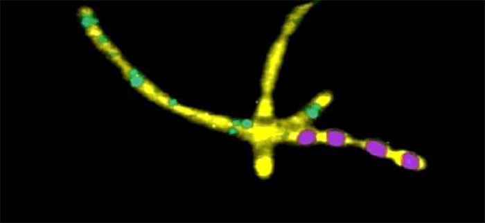
Confocal Raman image of Bacillus Cereus.
Working Principles
Confocal Raman Imaging

A Raman spectrum shows the energy shift of the excitation light (laser) as a result of inelastic scattering by the molecules in a sample. The excitation light excites or annihilates vibrations of the chemical bonds within the molecules which results in an energy shift of the photon scattered from this molecule. Different chemical species consist of different atoms and bonds, so each molecule can be easily identified by its unique Raman spectrum. As only molecular vibrations are excited (or annihilated), Raman spectroscopy is a nondestructive technique. In Raman imaging the Raman spectra are collected with a highthroughput confocal microscope/Raman spectrometercombination. A high-sensitivity CCD camera connected to a powerful computer and software system is used to detect the Raman signal. With specialized software tools the imaging capabilities can be expanded even further. For example, it is possible to generate images by integrating over selected spectral areas, determining the peak width, peak position or by even more sophisticated procedures such as the fitting of complete spectra or cluster analysis.

Atomic Force Microscopy (AFM)

In Atomic Force Microscopy the sample is scanned under the tip using a piezo-driven scanning-stage and the topography is displayed as an image. Atomic Force Microscopy provides spatial information parallel and perpendicular to the surface with resolution in the nm range. In addition to topographic high-resolution information, local material properties such as adhesion and stiffness can be investigated by analyzing the tip-sample interaction forces.

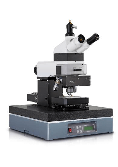
Raman-AFM Microscope alpha300 RA
>> Download the full Application Note as PDF

Newsletters

Receive the latest pathologist news, personalities, education, and career development – weekly to your inbox.

Newsletter Signup Image

Explore More in Pathology

Dive deeper into the world of pathology. Explore the latest articles, case studies, expert insights, and groundbreaking research.

False

Advertisement

Recommended

False

Related Content

Dioxin determination on air filter samples

January 12, 2016

This application note describes the pressurized solvent extraction of dioxins and furans from air filter samples (PUF and QFF) using the SpeedExtractor E-914...

Vitamin D Level Determination in Serum

February 2, 2016

Vitamin D3 (cholecalciferol) is essential for the human body...

Determination of the acid value, hydroxyl value, and isocyanates in raw materials for the fabrication of plastics by automatic potentiometric titration

February 8, 2016

The determination of the acid value, the hydroxyl value, and the isocyanates plays an important part in the analysis of raw materials for plastics...

Water determination in various plastics

February 8, 2016

The presence of excessive water in plastics adversely affects the performance of polymeric goods which is why water determination is of crucial importance...

False

The Analytical Scientist
Subscribe

About

  • About Us
  • Work at Conexiant Europe
  • Terms and Conditions
  • Privacy Policy
  • Advertise With Us
  • Contact Us

Copyright © 2025 Texere Publishing Limited (trading as Conexiant), with registered number 08113419 whose registered office is at Booths No. 1, Booths Park, Chelford Road, Knutsford, England, WA16 8GS.