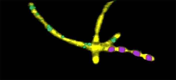
From measurements in liquids, to solid samples and soft tissues in life science research various samples are analysed on a regular basis. Through their convenient handling and versatile analysing possibilities all WITec microscope systems are particularly well-suited for life science applications: The flexible WITec imaging tools provide the opportunity to adjust the imaging technique to varying requirements and the powerful WITec software enables the comprehensive evaluation of the acquired data and the generation of depth profiles and 3D images.

 Confocal Raman image of Bacillus Cereus.
Working Principles
Confocal Raman Imaging
A Raman spectrum shows the energy shift of the excitation light (laser) as a result of inelastic scattering by the molecules in a sample. The excitation light excites or annihilates vibrations of the chemical bonds within the molecules which results in an energy shift of the photon scattered from this molecule. Different chemical species consist of different atoms and bonds, so each molecule can be easily identified by its unique Raman spectrum. As only molecular vibrations are excited (or annihilated), Raman spectroscopy is a nondestructive technique. In Raman imaging the Raman spectra are collected with a highthroughput confocal microscope/Raman spectrometercombination. A high-sensitivity CCD camera connected to a powerful computer and software system is used to detect the Raman signal. With specialized software tools the imaging capabilities can be expanded even further. For example, it is possible to generate images by integrating over selected spectral areas, determining the peak width, peak position or by even more sophisticated procedures such as the fitting of complete spectra or cluster analysis.
Atomic Force Microscopy (AFM)
In Atomic Force Microscopy the sample is scanned under the tip using a piezo-driven scanning-stage and the topography is displayed as an image. Atomic Force Microscopy provides spatial information parallel and perpendicular to the surface with resolution in the nm range. In addition to topographic high-resolution information, local material properties such as adhesion and stiffness can be investigated by analyzing the tip-sample interaction forces.
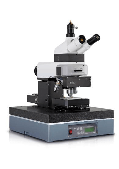
 Raman-AFM Microscope alpha300 RA


