Conexiant
Login
  • The Analytical Scientist
  • The Cannabis Scientist
  • The Medicine Maker
  • The Ophthalmologist
  • The Pathologist
  • The Traditional Scientist
The Analytical Scientist
  • Explore

    Explore

    • Latest
    • News & Research
    • Trends & Challenges
    • Keynote Interviews
    • Opinion & Personal Narratives
    • Product Profiles
    • App Notes

    Featured Topics

    • Mass Spectrometry
    • Chromatography
    • Spectroscopy

    Issues

    • Latest Issue
    • Archive
  • Topics

    Techniques & Tools

    • Mass Spectrometry
    • Chromatography
    • Spectroscopy
    • Microscopy
    • Sensors
    • Data and AI

    • View All Topics

    Applications & Fields

    • Clinical
    • Environmental
    • Food, Beverage & Agriculture
    • Pharma and Biopharma
    • Omics
    • Forensics
  • People & Profiles

    People & Profiles

    • Power List
    • Voices in the Community
    • Sitting Down With
    • Authors & Contributors
  • Business & Education

    Business & Education

    • Innovation
    • Business & Entrepreneurship
    • Career Pathways
  • Events
    • Live Events
    • Webinars
  • Multimedia
    • Video
Subscribe
Subscribe

False

The Analytical Scientist / App Notes / 2024 / Correlative Confocal Raman Microscopy for 2D Materials Investigation

Correlative Confocal Raman Microscopy for 2D Materials Investigation

06/21/2024

Share

Featured Image

The Raman principle 
 

The Raman effect is based on the inelastic scattering of light by the molecules of gaseous, liquid or solid materials. The interaction of a molecule with photons causes vibrations of its chemical bonds, leading to specific energy shifts in the scattered light. Thus, any given chemical compound produces a particular Raman spectrum when excited and can be easily identified by this individual “fingerprint.” Raman spectroscopy is a well- established, label-free and non- destructive method for analyzing the molecular composition of a sample.

Raman imaging 
 

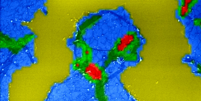
In Raman imaging, a confocal microscope is combined with a spectrometer and a Raman spectrum is recorded at every image pixel. The resulting Raman image visualizes the distribution of the sample’s compounds. Due to the high confocality of WITec Raman systems, volume scans and 3D images can also be generated. 

No need for compromises 
 

The Raman effect is extremely weak, so every Raman photon is important for imaging. Therefore WITec Raman imaging systems combine an exceptionally sensitive confocal microscope with an ultra-high throughput spectrometer (UHTS). Precise adjustment of all optical and mechanical elements guarantees the highest resolution, outstanding speed and extraordinary sensitivity – simultaneously! This optimization allows the detection of Raman signals of even weak Raman scatterers and extremely low material concentrations or volumes with the lowest excitation energy levels. This is an unrivaled advantage of WITec systems.
 

>> Download the Application Note as a PDF

Newsletters

Receive the latest analytical scientist news, personalities, education, and career development – weekly to your inbox.

Newsletter Signup Image

Explore More in Analytical Science

Dive deeper into the analytical science. Explore the latest articles, case studies, expert insights, and groundbreaking research.

False

Advertisement

Recommended

False

Related Content

NDIR gas sensing, Improve your detector design

January 23, 2024

This article explains how to detect certain gases ...

Charge heterogeneity characterisation of an IgG4-based mAb using AEX coupled to MS

January 26, 2024

Cation exchange chromatography (CEX) is very well suited for characterising the charge heterogeneity of biomolecules...

Improving Feature Detection and Putative Identification for Tissue Imaging Applications

January 29, 2024

HRMS DESI Imaging with the SELECT SERIES MRT Mass ...

Enhancing Biotransformation Identification Efficiency Using LC-MS Fine Isotope Structure Produced With Multi Reflecting Time-of-Flight MS

January 29, 2024

HRMS coupled with LC is an ideal analytical tool f...

False

The Analytical Scientist
Subscribe

About

  • About Us
  • Work at Conexiant Europe
  • Terms and Conditions
  • Privacy Policy
  • Advertise With Us
  • Contact Us

Copyright © 2025 Texere Publishing Limited (trading as Conexiant), with registered number 08113419 whose registered office is at Booths No. 1, Booths Park, Chelford Road, Knutsford, England, WA16 8GS.