Conexiant
Login
  • The Analytical Scientist
  • The Cannabis Scientist
  • The Medicine Maker
  • The Ophthalmologist
  • The Pathologist
  • The Traditional Scientist
The Analytical Scientist
  • Explore

    Explore

    • Latest
    • News & Research
    • Trends & Challenges
    • Keynote Interviews
    • Opinion & Personal Narratives
    • Product Profiles
    • App Notes

    Featured Topics

    • Mass Spectrometry
    • Chromatography
    • Spectroscopy

    Issues

    • Latest Issue
    • Archive
  • Topics

    Techniques & Tools

    • Mass Spectrometry
    • Chromatography
    • Spectroscopy
    • Microscopy
    • Sensors
    • Data and AI

    • View All Topics

    Applications & Fields

    • Clinical
    • Environmental
    • Food, Beverage & Agriculture
    • Pharma and Biopharma
    • Omics
    • Forensics
  • People & Profiles

    People & Profiles

    • Power List
    • Voices in the Community
    • Sitting Down With
    • Authors & Contributors
  • Business & Education

    Business & Education

    • Innovation
    • Business & Entrepreneurship
    • Career Pathways
  • Events
    • Live Events
    • Webinars
  • Multimedia
    • Video
Subscribe
Subscribe

False

The Analytical Scientist / App Notes / 2020 / Best practice examples from various fields of application

Best practice examples from various fields of application

06/19/2020

Share

Featured Image

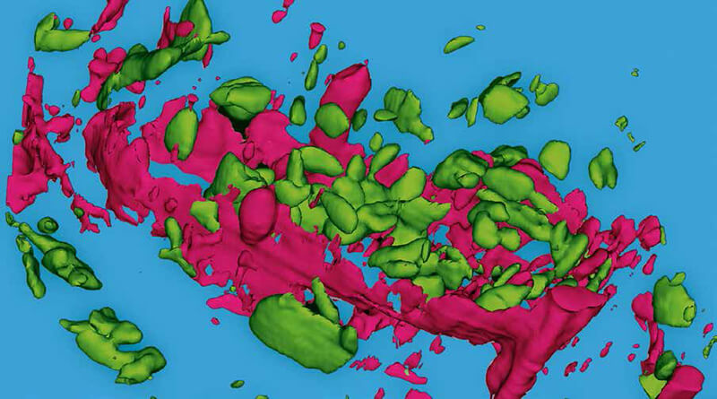
Confocal Raman microscopy is a powerful technique for highresolution, non-destructive and label-free chemical analysis. It is well-suited for three-dimensional investigation of samples from various fields of application.

Confocal Raman imaging is a microscopy technique for identifying and imaging of chemical and molecular compounds. This non-destructive, non-invasive and label-free method is used in various fields of application such as nanotechnology, materials and surface research, geological, environmental and life sciences and pharmaceutics.

The Raman effect (or Raman scattering) is based on the inelastic scattering of light interacting with molecules in a sample. This interaction causes vibrations in the chemical bonds, leading to a specific energy shift in the scattered light that is visible in its spectrum. The Raman spectrum is as unique for each chemical compound as a fingerprint and can be detected and identified by Raman spectroscopy.

For confocal Raman imaging a Raman spectrometer is combined with a confocal optical microscope. The microscope facilitates morphological characterization and establishes the spatial distribution of chemical components within a sample. High-resolution confocal Raman systems acquire the information of a complete Raman spectrum at every image pixel and achieve a lateral resolution at the diffraction limit. The confocal microscope setup is furthermore distinguished by a high signal-to-noise ratio and enables the generation of 3D Raman images and depth profiles. Additional sample characteristics such as the relative amount of a specific component, stress and strain states, or crystallinity can be further analyzed and imaged.
>> Download the Full Application Note as a PDF​​​​​​​

Newsletters

Receive the latest analytical scientist news, personalities, education, and career development – weekly to your inbox.

Newsletter Signup Image

Explore More in Analytical Science

Dive deeper into the analytical science. Explore the latest articles, case studies, expert insights, and groundbreaking research.

False

Advertisement

Recommended

False

Related Content

Detection of residual pesticides on fruits and vegetables using Portability™ miniature mass spectrometer

June 9, 2020

Mass spectrometry can be now deployed for onsite pesticide screening in real time...

Routine monitoring of airborne VOCs using TD–GC×GC–TOF MS/FID

January 15, 2020

The identification and quantitation of volatile organic pollutants in air can be challenging, largely because of the complexity of the samples...

Quantifying trace odorants in water by GC–MS with trap-based preconcentration

January 20, 2020

Assessing SPME and high‑capacity sorptive extracti...

Using Self-Aware Agilent InfinityLab LC/MSD iQ to Measure Trace-Level Impurities in a Brand Versus Generic Medication

January 22, 2020

This study presents a method for comparison of related impurities in a brand and a generic over-the-counter (OTC) acetaminophen drug...

False

The Analytical Scientist
Subscribe

About

  • About Us
  • Work at Conexiant Europe
  • Terms and Conditions
  • Privacy Policy
  • Advertise With Us
  • Contact Us

Copyright © 2025 Texere Publishing Limited (trading as Conexiant), with registered number 08113419 whose registered office is at Booths No. 1, Booths Park, Chelford Road, Knutsford, England, WA16 8GS.