Conexiant
Login
  • The Analytical Scientist
  • The Cannabis Scientist
  • The Medicine Maker
  • The Ophthalmologist
  • The Pathologist
  • The Traditional Scientist
The Analytical Scientist
  • Explore

    Explore

    • Latest
    • News & Research
    • Trends & Challenges
    • Keynote Interviews
    • Opinion & Personal Narratives
    • Product Profiles
    • App Notes

    Featured Topics

    • Mass Spectrometry
    • Chromatography
    • Spectroscopy

    Issues

    • Latest Issue
    • Archive
  • Topics

    Techniques & Tools

    • Mass Spectrometry
    • Chromatography
    • Spectroscopy
    • Microscopy
    • Sensors
    • Data and AI

    • View All Topics

    Applications & Fields

    • Clinical
    • Environmental
    • Food, Beverage & Agriculture
    • Pharma and Biopharma
    • Omics
    • Forensics
  • People & Profiles

    People & Profiles

    • Power List
    • Voices in the Community
    • Sitting Down With
    • Authors & Contributors
  • Business & Education

    Business & Education

    • Innovation
    • Business & Entrepreneurship
    • Career Pathways
  • Events
    • Live Events
    • Webinars
  • Multimedia
    • Video
Subscribe
Subscribe

False

The Analytical Scientist / App Notes / 2021 / Food Analysis with Confocal Raman Microscopy

Food Analysis with Confocal Raman Microscopy

09/21/2021

Share

Featured Image

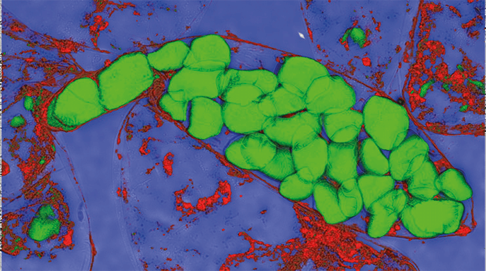
Confocal Raman microscopy is a powerful tool for visualizing the che- mical composition of heterogeneous samples on the sub-micrometer scale. This application note demonstrates the utility of Raman imaging for characterizing various food samples such as honey, chocolate and fat- spreads, leading to a comprehensive understanding of the products and the production processes.

The Raman principle

The Raman effect is based on the inelastic scattering of light by the molecules of gaseous, liquid or solid materials. The interaction of a molecule with photons causes vibrations of its chemical bonds, leading to specific energy shifts in the scattered light. Thus, any given chemical compound produces a particular Raman spectrum when excited and can be easily identified by this individual “fingerprint.” Raman spectroscopy is a well-established, label-free and non-destructive method for analyzing the molecular composition of a sample.

Raman imaging

In Raman imaging, a confocal microscope is combined with a spectrometer and a Raman spectrum is recorded at every image pixel. The resulting Raman image visualizes the distribution of the sample’s compounds. Due to the high confocality of WITec Raman systems, volume scans and 3D images can also be generated.

No need for compromises

The Raman effect is extremely weak, so every Raman photon is important for imaging. Therefore WITec Raman imaging systems combine an exceptionally sensitive confocal microscope with an ultra-high throughput spectrometer (UHTS). Precise adjustment of all optical and mechanical elements guarantees the highest resolution, outstanding speed and extraordinary sensitivity - simultaneously! This optimization allows the detection of Raman signals of even weak Raman scatterers and extremely low material concentrations or volumes with the lowest excitation energy levels. This is an unrivaled advantage of WITec systems.

>> Download the Full Application Note as a PDF

Newsletters

Receive the latest pathologist news, personalities, education, and career development – weekly to your inbox.

Newsletter Signup Image

Explore More in Pathology

Dive deeper into the world of pathology. Explore the latest articles, case studies, expert insights, and groundbreaking research.

False

Advertisement

Recommended

False

Related Content

Removing User Bias from Structure Verification by NMR

November 15, 2021

Chemical structure verification by NMR is one of t...

Simultaneous Determination of Eight Nitrosamine Impurities in Metformin Extended-Release Tablets Using the Agilent 6470 Triple Quadrupole LC/MS

November 15, 2021

Detection of regulated genotoxic impurities from t...

HIGH RESOLUTION MULTI-REFLECTING TIME-OF-FLIGHT MASS ANALYZER WITH FOLDED FLIGHT PATH®

November 15, 2021

How high does resolving power need to be?

Biomarker discovery
Biomarker discovery

November 16, 2021

Essential guide to analysing VOCs in breath and other biological samples...

False

The Analytical Scientist
Subscribe

About

  • About Us
  • Work at Conexiant Europe
  • Terms and Conditions
  • Privacy Policy
  • Advertise With Us
  • Contact Us

Copyright © 2025 Texere Publishing Limited (trading as Conexiant), with registered number 08113419 whose registered office is at Booths No. 1, Booths Park, Chelford Road, Knutsford, England, WA16 8GS.