I am a postdoctoral researcher, splitting my time between the University of Amsterdam and the Rijksmuseum. I am trained as a chemist, and work on fundamental questions relating to oil paint degradation.
Model behavior
I do not often carry out analysis of real artworks. Instead, I design and create model systems that mimic the molecular structure or chemical reactivity of real aged oil paint, while still being suitable for a particular type of chemical analysis. In this way, it is possible to study in great detail how the materials in oil paint react with each other, and how fast these processes occur under certain conditions. Eventually, all this knowledge will be used to understand how current methods of oil paint conservation and restoration are associated with the risk of future degradation, and how we can minimize that risk.
Right now, it is very difficult to make a reasonable quantitative estimate of the risks involved with current practice for the conservation and restoration of paintings. How sensitive is a painting to changes in humidity or temperature, or how much impact does treatment with solvents have on the stability of a painting? While the extensive experience of conservators is a very good starting point, for better risk assessment we need to first understand the chemical and physical processes behind paint degradation. Secondly, we need to measure how the rate of paint degradation is affected by environmental conditions or restoration treatments.

We are still in the early stages of this research, unraveling the immensely complex set of chemical reactions and diffusion processes that occur in aged oil paint. However, we have already uncovered potential markers that can help determine how a painting is likely to change in the future. We are in constant discussion with the conservators of the Rijksmuseum and other museums around the world to test our ideas and share our findings. Together, we work to extend the lifetime of oil paintings for the benefit of future generations.
Imitating art
The model systems we design must strike the right balance of being tunable (so we have control over relevant parameters), while still yielding information that is useful for understanding real-world degradation. The formation of metal soaps in oil paint is a good example. If you mix a lead- or zinc-containing pigment with oil and let it age for a few months under humid conditions, metal soaps (complexes of metal ions and saturated fatty acids) will spontaneously form. However, if you want to understand what is really going on, you need to design systems in which you can break down the process into its constituent steps: the reaction between pigments and relevant functional groups of the oil, the formation of fatty acids in the oil, the reaction between fatty acids and the released metal ions to form metal soaps, and the crystallization of the metal soap end products.
In real paintings these processes operate on vastly different timescales, so creative solutions are usually required to make them happen on a scale we can analyze. In addition, reactions in real paintings take place in an insoluble cross-linked polymer matrix, where most reactivity is diffusion-limited. To carry out analyses like infrared (IR) spectroscopy, MS or XRD, the relevant reactions sometimes need to be replicated in different environments to allow measurement.
Light at the museum
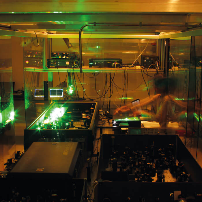
Our main workhorse is IR spectroscopy, in all its forms. With ATRFTIR spectroscopy we can identify certain pigments and degradation products (like metal soaps) in oil paints or oil paint models, and follow the changes in chemical composition over time in situ while we expose samples to humidity, temperature or reactant solutions. We then apply custom-made spectral processing algorithms to the datasets to obtain concentration profiles, diffusion constants and rate constants. Recently, we have started applying the same approach to IR microscopy measurements, so we can follow diffusion of chemical species and their reactivity over time with micrometer spatial resolution. The main advantage of IR spectroscopy is that it is easy to use and relatively easy to interpret. We also make use of complementary techniques like XRD, DSC, SAXS, NMR, and even femtosecond pump-probe 2D-IR spectroscopy, to help us understand the molecular structure of our materials.
I see the field changing, with ever more data being collected on every painting during conservation treatment. On the horizon, we are seeing automated hyperspectral imaging systems being developed that can be used to identify pigments, and XRDimaging systems that can be used to map crystalline phases with sub-millimeter spatial resolution in a painting.
As a consequence, major advances are needed in processing, correlating, matching and interpreting data. Machine learning algorithms and other forms of automation will be necessary, but it is crucial to keep thinking about the questions you really want to answer. What do we really need to know about a painting, and is that information worth the financial and time investment? How can we extract meaningful information out of a mountain of data? These questions are going to become more and more important in the coming years.


