Essential Reading
Freeze Frame
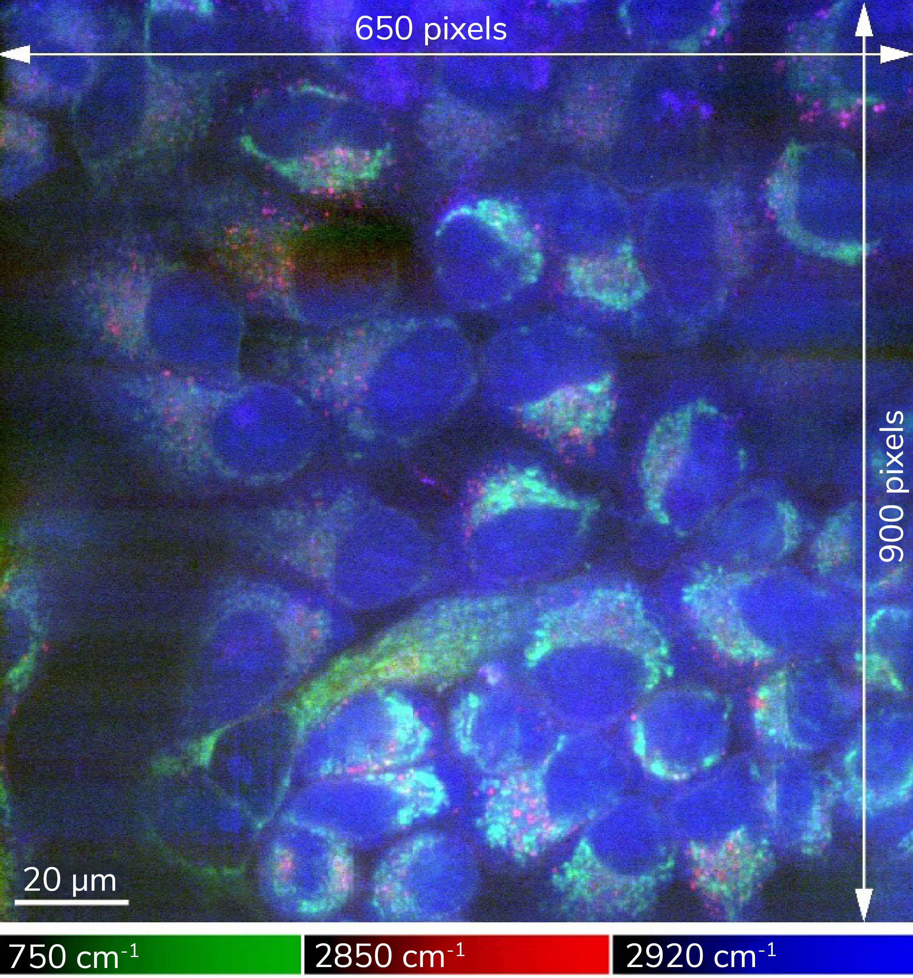
Raman microscopy has emerged as a promising, non-destructive tool for imaging the distribution of molecules in biological samples, allowing researchers to, for example, visualize cell dynamics and drug effects. The main challenge, however, lies in extracting a strong, reliable signal from the biomolecular din. A solution suggested by research from Osaka University, Japan, is to “cryofix” (that is, freeze) the samples, thereby reducing molecular motion over long acquisition times to produce sharper chemical images – eight times sharper, the researchers found.
"By imaging frozen samples that were unable to move, we could use longer exposure times without damaging the samples,” said lead author Kenta Mizushima in a press release. “This led to high signals compared with the background, high resolution, and larger fields of view."
Straight to the Point
Researchers from China have developed a miniaturized, all-fiber photoacoustic spectrometer (FPAS); its key components, i.e. the photoacoustic gas cell and optical microphone, have been integrated into a tip with a diameter of just 125 μm. Despite its small size, the new system is almost as sensitive as larger, traditional laboratory spectrometers – reaching detection limits for acetylene as low as 9 ppb. The researchers believe the new system could be used for real-time and in situ trace gas measurement in various fields, such as intravascular blood gas monitoring, lithium-ion battery health assessments, and remote detection of explosive gas leakage.

Raman image of rapidly frozen HeLa cells with high signal-to-noise ratio and large field-of-view. The image acquisition time was 10 hours. The distribution of Raman signals from cytochromes (750 cm⁻¹), lipids (2850 cm⁻¹), proteins (2920 cm⁻¹), are indicated in green, red, and blue, respectively.
Credit: Sci. Adv. 10, eadn0110 (2024)
Also in the news…
Researchers report mid-infrared photothermal plasmonic scattering (MIP-PS) spectroscopy with ultrahigh sensitivity to detect a trace amount of small molecules – heralding potential in bond-selective biosensing and bioimaging. Link
FTIR spectroscopy combined with machine learning detects glioblastoma G4 and meningiomas in tissue samples with an accuracy and specificity of more than 90 percent. Link
Researchers successfully reproduce Ziatdinov et al.’s AtomAI – a comprehensive Python library designed for a wide range of materials imaging tasks – research; “We believe that AtomAI holds significant potential for the microscopy and spectroscopy communities,” the researchers conclude. Link
Vibrational fiber photometry used for non-invasive label-free monitoring of the biomolecular content of deep regions of the mouse brain in vivo through spontaneous Raman spectroscopy – with application to fundamental and preclinical investigations of the brain and other organs. Link
Researchers use single-molecule AFM-based force spectroscopy to investigate how the SARS-CoV-2 spike protein interacts with host cell membranes, finding that cholesterol depletion significantly reduces viral infectivity; the results suggest that targeting the disulfide bridge could provide a therapeutic strategy against infection. Link
Study demonstrates the potential of Raman Spectroscopy and multimodal imaging to interrogate cartilage tissue and provides insight into the chemical and structural composition of its different layers with significant implications for osteoarthritis diagnosis. Link


