With confocal Raman imaging the molecules of a sample can be chemically identified and their distribution can be imaged three-dimensionally. These benefits are gaining more and more recognition in biological and medical research and confocal Raman microscopy (CRM) is becoming a more frequently used method for answering crucial questions in life sciences. CRM is used for measurements in liquids and live cell imaging, solid samples and soft tissues. Through various examples this application note describes possible uses of confocal Raman microscopy and correlative techniques.

Working principles
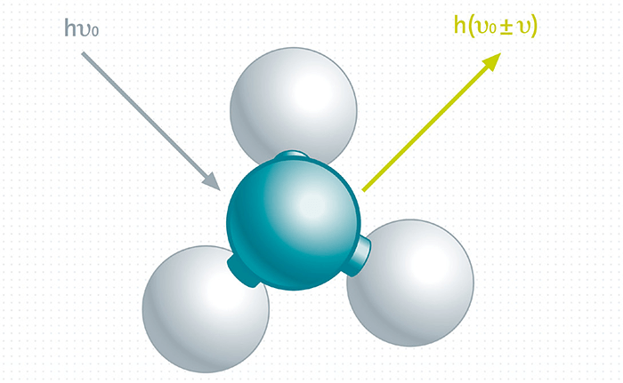
Confocal Raman Imaging The Raman effect is based on light interacting with the chemical bonds within a sample. This causes a specific energy shift in the backscattered light which appears in a unique Raman spectrum through which the molecular components of a sample can be detected. The confocal Raman imaging technique combines Raman spectroscopy with a confocal microscope. This allows the spatial distribution of the chemical components within the sample to be imaged. High-resolution confocal Raman microscopes acquire a complete Raman spectrum at every image pixel and achieve a lateral resolution limited only by diffraction (circa λ/2 of the excitation wavelength). A confocal microscope setup is furthermore characterized by an excellent depth resolution and facilitates the generation of 3D Raman images and depth profiles. Confocal Raman microscopy can be coupled with correlative microscopy techniques such as fluorescence microscopy, electron microscopy and atomic force microscopy.
>> Download the full Application Note as PDF
Newsletters
Receive the latest pathologist news, personalities, education, and career development – weekly to your inbox.