Introduction
Development, production and quality control in the pharmaceutical industry requires efficient and reliable control mechanisms to ensure the safety and therapeutic effect of the final products. These products can vary widely in composition and application. Several of their properties can be difficult to study with conventional techniques due to the inability of these methods to chemically differentiate materials with sufficient spatial resolution and without damage or staining. Therefore analyzing methods that provide both comprehensive chemical characterization and the flexibility to adjust the method to the investigated specimen are preferred in pharmaceutical research.

Confocal Raman Microscopy (CRM) is a well-established and widely-used spectroscopic method for the investigation of the chemical composition of a sample. In pharmaceutical research, CRM can be used to probe the distribution of components within formulations, to characterize the homogeneity of pharmaceutical samples, to determine the state of drug substances and excipients and to characterize contaminants and foreign particulates [1]. The information obtained by CRM is also useful for drug substance design, for the development of solid and liquid formulations, as a tool for process analytics and for patent infringement and counterfeit analysis [2, 3, 4]. Being a non-destructive method it is possible to combine CRM with other imaging techniques such as Atomic Force Microscopy (AFM), Scanning Electron Microscopy (SEM) or fluorescence analysis.
The Aim of the Study
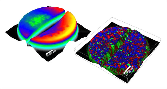
Based on various samples the article will demonstrate how CRM can be beneficially applied in the pharmaceutical field. With high-resolution, large-area Raman images, topographic 3D Raman images, depth-profiles and correlative Raman-AFM images the analyzing capabilities of CRM and its correlative techniques will be shown. Furthermore an example of a statistical quantitative evaluation of a spectral Raman dataset will be given.
>> Download the full Application Note as PDF
Newsletters
Receive the latest pathologist news, personalities, education, and career development – weekly to your inbox.