Subscribe to Newsletter
Fields & Applications Pharma & Biopharma

A Picture Paints a Thousand… New Therapies?

Cell painting is a high-content profiling technology that uses up to six fluorescent dyes to visualize specific cellular components at the single-cell level. The assay essentially “paints” images of various components of the cell, including the nucleus, mitochondria, endoplasmic reticulum, cytoskeleton, and more. After microscopic images are captured, image analysis software converts them to data by extracting various measures of cellular morphology, called features. The results allow researchers to understand the effects of perturbagens, such as chemical compounds or genes, on the behavior of cells, feeding drug discovery and the characterization of bioactive molecules. 

Here, we speak with Angeline Lim, Senior Scientist at Molecular Devices, and David Egan, co-founder and CEO at Core Life Analytics, about a collaboration focused on using AI to make cell painting faster and more efficient.

What are the current limitations of cell painting? 
 

Cell painting workflows often prove time- and labor-intensive – a full screen may take several days to complete and generate massive amounts of data. Next, researchers face the challenge of extracting meaningful information from the resulting data, which can be overwhelming – either demanding the expertise of a data scientist or – in a worst-case scenario – wasting valuable data simply because of the lack of tools. 

How can AI-enabled technologies help? 
 

Overcoming the aforementioned hurdles requires additional computational tools. In the past, only a team of statisticians, data scientists, and software developers was capable of analyzing cell painting data with any reasonable and timely success. Now, AI can quickly facilitate the analysis of large image datasets. In addition to machine learning-enabled software that handles data extraction from images, AI-enabled tools such as StratoMineR – a data analytics software from Core Life Analytics – can also help scientists iteratively mine data for information without the help of a data scientist.

Could you share more details about your collaboration?
 

At Molecular Devices, our customers are largely biologists in the life sciences field – not data scientists – who seek us out for guidance around analyzing large datasets coming from their cell painting assays performed with our high-content cellular imaging systems. The cloud-powered data analytics solution from Core Life Analytics, called StratoMineR, offers users a self-guided workflow that enables researchers to easily derive meaning from their high-content data.

Our collaboration provides more biologists with the AI-enabled tools that deliver advanced data mining and analysis – reducing the dependency on data science expertise. 

And how might this benefit drug discovery?  
 

The democratization of data science with AI-enabled analytic software enables data-driven decision making. It has enormous potential for speeding up the drug discovery process and reducing failure rates.
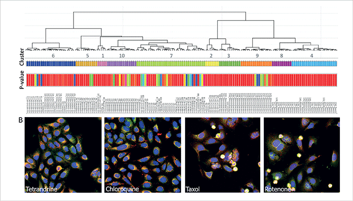
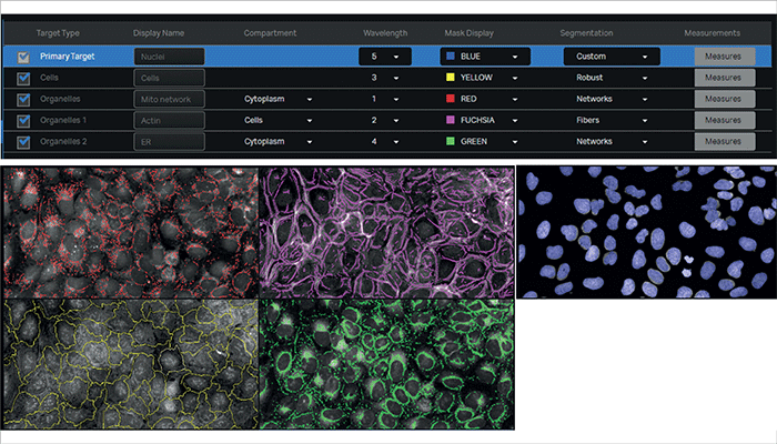
Figure 1. Cluster analysis. A) A dendrogram that represents hierarchical relationships is shown. Wells belonging to the same cluster (numbered) are represented by colored bars. P-values based on the distance score are shown for each well. Note that cluster 9 consists of only staurosporine treated cells, whereas cluster 10 consists of only etoposide treated cells. B) Examples of compound-treated cells belonging to some of the clusters are shown. Cluster 5 consists of tetrandrine- and chloroquine-treated cells. Note the increased number of ER punctae in both wells. Cluster 4 consists of rotenone- and paclitaxel-treated cells. Note the presence of blebbing in some of the cells belonging in these wells, suggesting cytotoxic effects.

Figure 2. Feature extraction in IN Carta software. A) An analysis protocol was created in the IN Carta software to segment the various cellular structures. Here, we used the built-in nuclei model to achieve robust segmentation of nuclei across all treatments. Other cellular features such as cytoplasm compartment, actin filament network, ER, and mitochondria were also segmented.

Angeline Lim supports scientific, technical, and applications initiatives for Molecular Devices’ portfolio of ImageXpress high-content imaging systems and David Egan co-developed Core Life Analytics’ StratoMineR platform with his co-founder Wienand Omta to help biologists independently analyze complex datasets.

Receive content, products, events as well as relevant industry updates from The Analytical Scientist and its sponsors.
Stay up to date with our other newsletters and sponsors information, tailored specifically to the fields you are interested in

When you click “Subscribe” we will email you a link, which you must click to verify the email address above and activate your subscription. If you do not receive this email, please contact us at [email protected].
If you wish to unsubscribe, you can update your preferences at any point.

About the Author
James Strachan

Over the course of my Biomedical Sciences degree it dawned on me that my goal of becoming a scientist didn’t quite mesh with my lack of affinity for lab work. Thinking on my decision to pursue biology rather than English at age 15 – despite an aptitude for the latter – I realized that science writing was a way to combine what I loved with what I was good at.

From there I set out to gather as much freelancing experience as I could, spending 2 years developing scientific content for International Innovation, before completing an MSc in Science Communication. After gaining invaluable experience in supporting the communications efforts of CERN and IN-PART, I joined Texere – where I am focused on producing consistently engaging, cutting-edge and innovative content for our specialist audiences around the world.


Related Application Notes
Charge heterogeneity analysis of an acidic protein and identification of its proteoforms using a streamlined icIEF-UV/MS workflow

| Contributed by SCIEX

Site-specific differentiation of hydroxyproline isomers using electron activated dissociation (EAD)

| Contributed by SCIEX

Charge variant analysis of antibody-drug conjugates using an icIEF-UV/MS workflow

| Contributed by SCIEX

Related Product Profiles
Higher Peaks – Clearly.

| Contributed by Shimadzu Europa

Compact with countless benefits

| Contributed by Shimadzu Europa

The fine Art of Method Development

| Contributed by Shimadzu Europa

Register to The Analytical Scientist

Register to access our FREE online portfolio, request the magazine in print and manage your preferences.

You will benefit from:
  • Unlimited access to ALL articles
  • News, interviews & opinions from leading industry experts
  • Receive print (and PDF) copies of The Analytical Scientist magazine

Register