Subscribe to Newsletter
Fields & Applications Mass Spectrometry, Clinical, Forensics

Collaborating for the Clinical Win

Is there a common theme to your career?

Change and passion. I always told myself that every ten years I would do something different. I was trained as a physicist, became a professor in chemistry, and now I am working in a molecular imaging institute housed in a medical department. I stay enthused about what I am doing by making it worthwhile – and changing my environment and research topics helps to keep my passion for science alive.

So, what motivates you?

Curiosity, enthusiasm, passion – wanting to be an explorer. I’m very lucky to have been able to set up this wonderful institute within the University of Maastricht. A collaborator of ours said to me recently, “I feel like a kid in a candy store!” And that’s exactly the environment that we wanted to create – it encourages people to do great analytical science and great molecular imaging. Here, I get to define the questions I’d like to ask and the best tools for answering those questions, and so explore the world around me. How much better does it get?

Tell us about your current research...

Our major focus is using molecular imaging based on mass spectrometry to assess the molecular content of tissues, so we can provide clinicians and medical researchers with feedback on the cellular phenotype. Say a surgeon operates on a patient and removes a tumor; it’s sent to a pathologist, who takes a section for hemotoxylin–eosin staining and inspection, and we take an adjacent section for mass spec imaging analysis. Half an hour later, we aim to provide the surgeon and pathologist with our findings and see how they match. In a second clinical research project – intraoperative diagnostics – we are analyzing the smoke from laser surgery to give surgeons the information they need in real time. Most of our research is biomedical, but we also use MS imaging to study new biomaterials, regenerative medicine, drug distribution and metabolism, forensics, and even historical paintings.

And recent findings?

In a study on cholangiocarcinoma we identified several peptides, proteins and lipids that distinguish transient neoplastic tissue (on its way to becoming a tumor) from full-blown tumor cells and healthy tissue. In other words, we can assess a single piece of tissue and define different cellular tissue phenotypes, which helps us to assess how clean the surgical margin of a tumor is; has the surgeon removed enough? Is there any cancerous or pre-cancerous tissue remaining?

Changing my environment and research topics helps to keep my passion for science alive.

How close are these tools to the clinic?

We’ve developed technology and methods for a number of diseases. The next step is validation – we need to work with large patient cohorts to make sure that the markers we have found in ten organs are stable and robust in 100 organs or 1,000 organs. That’s one reason the group is based in Maastricht, which gives us access to a dynamic clinical environment with a large volume of samples.

To establish a validated clinical diagnostic test, several major clinical studies and a lot of administrative and legal paperwork are needed. It’s difficult to predict how long it will take, but I hope that within 3-5 years some of these tests will be routinely available. For now, they are still in the research phase.

What is the role of analytical scientists in clinical collaborations?

Typically, the analytical scientist provides the technology and data needed to make a clinical assessment. The analytical scientist also needs to understand the questions or challenges faced by medical practitioners, and how new techniques could fit into the clinical workflow. They are the axle in the wheel – a project manager, analytical scientist and communicator all in one. As an analytical scientist, you are too often forced into the role of a service provider, and that’s not the way we work. Our ethos is CORE – collaborative, open research and education.

What’s the secret to successful collaboration?

Crucial to the success of this type of multidisciplinary research is a willingness to give something up to ultimately gain a lot more. Sometimes we generate great results, but rather than presenting them at an analytical science conference ourselves, we ask the surgeons on the team to present them at a surgery conference. We may give up a little visibility in our own community but, in the long term, we have a much bigger impact in the clinical field – where it really matters.

How important have your communication skills been in your career?

Absolutely crucial. Without good communication, it is impossible to start new projects or to motivate other researchers to move in the same direction. To my mind, communication is nothing more than showing how passionate you are about what you’re doing, how much fun you’re having while doing it, and how good the results are. It’s also important for me to showcase and emphasize the importance of the work of my (younger) colleagues, without whom I would be dead in the water.

What does the perfect team look like?

I believe in heterogeneous teams. My current group has a very international flavor and is 50 percent female – and that makes it a very culturally diverse group. Diversity in passions and interests is also crucial for the success of any team – we have specialists in everything from clinical science to bioinformatics. But they all share one thing: a passion for research. The enthusiasm of young researchers really helps with building such an institute. We aim to come up with new and fresh ideas, and that characterizes my staff: young, willing, enthusiastic, eager, and impassioned by their research.

What are your career highlights, so far?

From a research perspective, the best thing has been seeing how the ideas we had ten years ago are being realized in the clinical environment. I’ve always believed our work would make a difference for patients, and seeing that start to happen is wonderful. Talking about it at TEDxMaastricht recently was an absolute highlight for me, personally. Molecular structure plays a much bigger role in disease than we previously thought, and being able to visualize that with the technology we’ve developed is fantastic. It’s also great to have the chance to improve the instruments we all rely on, by collaborating with the companies who make them.

Receive content, products, events as well as relevant industry updates from The Analytical Scientist and its sponsors.
Stay up to date with our other newsletters and sponsors information, tailored specifically to the fields you are interested in

When you click “Subscribe” we will email you a link, which you must click to verify the email address above and activate your subscription. If you do not receive this email, please contact us at [email protected].
If you wish to unsubscribe, you can update your preferences at any point.

About the Author
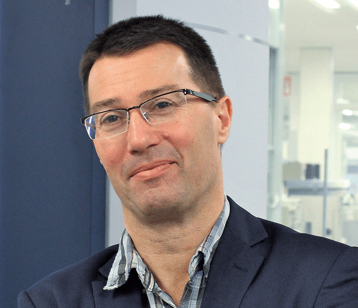
Ron M. A. Heeren

Director, M4I, and Distinguished Professor, University of Maastricht, Netherlands

Related Application Notes
Charge heterogeneity analysis of an acidic protein and identification of its proteoforms using a streamlined icIEF-UV/MS workflow

| Contributed by SCIEX

Site-specific differentiation of hydroxyproline isomers using electron activated dissociation (EAD)

| Contributed by SCIEX

High-Resolution Accurate Mass Library for Forensic Toxicology

| Contributed by Shimadzu

Related Product Profiles
ASMS 2024: Innovations Unveiled

Higher Peaks – Clearly.

| Contributed by Shimadzu Europa

Compact with countless benefits

| Contributed by Shimadzu Europa

Register to The Analytical Scientist

Register to access our FREE online portfolio, request the magazine in print and manage your preferences.

You will benefit from:
  • Unlimited access to ALL articles
  • News, interviews & opinions from leading industry experts
  • Receive print (and PDF) copies of The Analytical Scientist magazine

Register