Conexiant
Login
  • The Analytical Scientist
  • The Cannabis Scientist
  • The Medicine Maker
  • The Ophthalmologist
  • The Pathologist
  • The Traditional Scientist
The Analytical Scientist
  • Explore

    Explore

    • Latest
    • News & Research
    • Trends & Challenges
    • Keynote Interviews
    • Opinion & Personal Narratives
    • Product Profiles
    • App Notes

    Featured Topics

    • Mass Spectrometry
    • Chromatography
    • Spectroscopy

    Issues

    • Latest Issue
    • Archive
  • Topics

    Techniques & Tools

    • Mass Spectrometry
    • Chromatography
    • Spectroscopy
    • Microscopy
    • Sensors
    • Data and AI

    • View All Topics

    Applications & Fields

    • Clinical
    • Environmental
    • Food, Beverage & Agriculture
    • Pharma and Biopharma
    • Omics
    • Forensics
  • People & Profiles

    People & Profiles

    • Power List
    • Voices in the Community
    • Sitting Down With
    • Authors & Contributors
  • Business & Education

    Business & Education

    • Innovation
    • Business & Entrepreneurship
    • Career Pathways
  • Events
    • Live Events
    • Webinars
  • Multimedia
    • Video
Subscribe
Subscribe

False

The Analytical Scientist / Issues / 2022 / Sep / Deep Visual Proteomics
Clinical Omics Proteomics

Deep Visual Proteomics

How a new method could transform our understanding of cancer evolution – potentially exposing tumor vulnerabilities

By Margot Lespade 09/02/2022 1 min read

Share

A combination of artificial intelligence (AI) image analysis, laser microdissection, and ultra-high sensitivity mass spectrometry – dubbed “Deep Visual Proteomics” (DVP) – is allowing researchers to characterize thousands of proteins from single cell types while preserving spatial information (1).

“It is useful for a wide range of purposes – from better understanding the function of cell heterogeneity in basic research to diagnosing and treating disease processes in the context of clinical pathology,” says lead author of the study, Andreas Mund, who is an associate professor at the Novo Nordisk Foundation Center for Protein Research, University of Copenhagen.
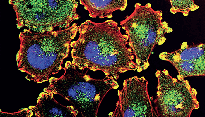
 

So how does it work? “DVP uses AI-based image analysis to identify cells or cellular regions of interest, which are then isolated by automated laser microdissection,” explains Mund. “We then use our single-cell proteomic sample preparation and ultra-high sensitivity mass spectrometry workflow. Based on the proteomes, we then assign cell functions to the imaging data. This uncovers a ‘microcosm in a single slide’ and reveals therapeutic vulnerabilities at different stages of tumor development.”

When examining the spatial distribution of a primary melanoma, DVP revealed how the proteome of normal melanocytes evolved as they become invasive melanoma cells within the same tissue slice. With this information gleaned from a single tissue slice, the researchers believe they will be able to reveal the mechanisms driving tumor development and expose new therapeutic targets.

DVP is clearly powerful, and Mund hopes others will benefit – in time: “We are now putting a lot of effort into making our tools user friendly and accessible to a wide range of researchers, but it is still an ongoing challenge.”

Newsletters

Receive the latest analytical science news, personalities, education, and career development – weekly to your inbox.

Newsletter Signup Image

References

  1. A Mund et al., Nature Biotechnology, 1 (2022). DOI: 10.1038/s41587-022-01302-5

About the Author(s)

Margot Lespade

More Articles by Margot Lespade

False

Advertisement

Recommended

False

Related Content

 This Week’s Mass Spec News
Clinical
This Week’s Mass Spec News

April 4, 2025

2 min read

Spit It Out
Clinical
Spit It Out

December 4, 2024

1 min read

Saliva-based PA-MS test detects paracetamol overdose in just 10 minutes

Let Me See That Brain
Clinical
Let Me See That Brain

December 9, 2024

1 min read

TRISCO sets a new standard for 3D RNA imaging, delivering high-resolution and uniform images to offer insights into brain function and anatomy

Meeting Mpox Where It’s At
Clinical
Meeting Mpox Where It’s At

December 17, 2024

1 min read

New optical biosensor offers mpox virus detection in 20 minutes

False

The Analytical Scientist
Subscribe

About

  • About Us
  • Work at Conexiant Europe
  • Terms and Conditions
  • Privacy Policy
  • Advertise With Us
  • Contact Us

Copyright © 2025 Texere Publishing Limited (trading as Conexiant), with registered number 08113419 whose registered office is at Booths No. 1, Booths Park, Chelford Road, Knutsford, England, WA16 8GS.