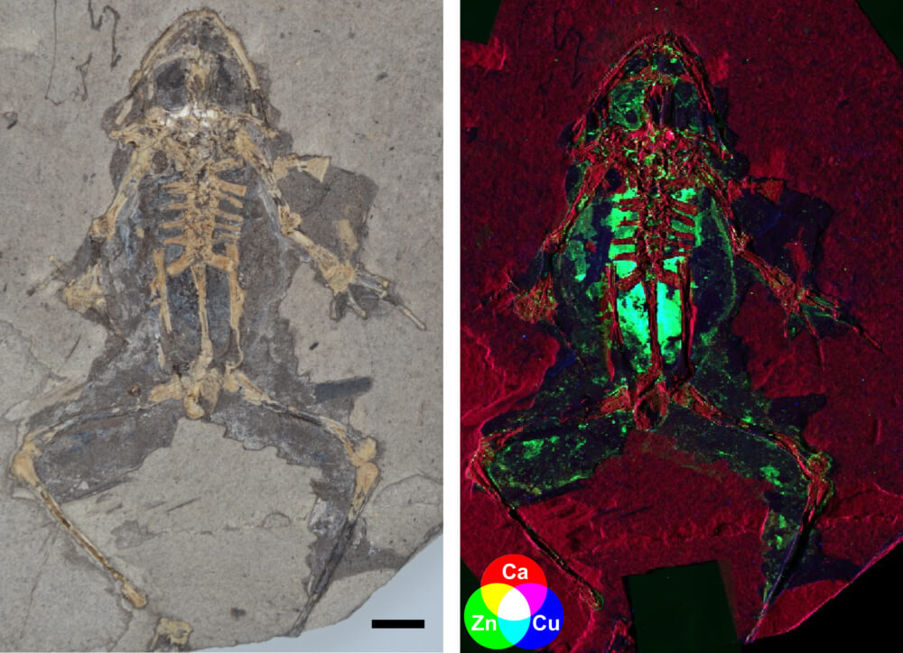
A combination of scanning electron microscopy and synchrotron rapid-scanning X-ray fluorescence has revealed the most anatomically detailed structures of fossilized frogs to date. Fossilized melanosomes – the cellular repositories of the pigment melanin, in addition to a variety of different metal ions – proved to be the perfect biomarker.
Credit: Fossil photograph copyright the Natural History Museum, London. X-ray fluorescence map copyright Valentina Rossi.
Reference: V Rossi et al., “Tissue-specific geometry and chemistry of modern and fossilized melanosomes reveal internal anatomy of extinct vertebrates”, Proc Natl Acad Sci U.S.A, [Epub ahead of print] (2019). DOI: 10.1073/pnas.1820285116
Would you like your photo featured in Image of the Month? Send it to charlotte.barker@texerepublishing.com
Newsletters
Receive the latest analytical science news, personalities, education, and career development – weekly to your inbox.