A study combining histological analysis and synchrotron X-ray microfluorescence has shed light on the causes of infant mortality in the Iron Age Iberian culture (8th–1st centuries BCE). By studying the teeth of infants buried within homes rather than cremated in necropolises, researchers have ruled out infanticide and ritual sacrifice as causes of death, concluding instead that most newborns died of natural causes, such as complications during birth or premature births.
The research analyzed 45 infant skeletal remains from five archaeological sites across northeastern Iberia, including Camp de les Lloses and Fortalesa dels Vilars d'Arbeca. The team were able to identify birth and death timings in infants by analyzing deciduous (primary) teeth, particularly focusing on the presence of the Neonatal Line (NNL), a growth line in teeth formed at birth.
To find out more about the research, we spoke with lead author Ani Martirosyan from The Autonomous University of Barcelona, Spain.

What was your inspiration for this work and the technique you developed?
The inspiration for this research stemmed from the ongoing debates surrounding the burials of infants in domestic and productive areas, a practice that has been observed across Europe since the Neolithic period and is particularly prevalent during the Iberian protohistoric period. These burials have often been interpreted as cases of infanticide or sacrifice, yet these claims lacked definitive proof. The aim of our work was to re-examine these interpretations by applying dental histology, focusing specifically on the analysis of the neonatal line in teeth.
The technique was inspired by previous studies that highlighted the usefulness of dental histology for identifying the neonatal line in different populations. Earlier research demonstrated that enamel growth patterns, particularly the formation of the neonatal line, could provide critical insights into whether an infant survived birth. The neonatal line forms in response to the physiological stress caused by the transition from intrauterine to extrauterine life. Studies conducted by researchers such as Antoine et al. (2009), Nava et al. (2017), Dean et al. (2014), and Mahoney (2015) were particularly influential, as they showed that by counting the daily increments in enamel, it was possible to accurately distinguish between stillbirths and live births. Their work served as a foundation for our approach, allowing us to use this technique to determine the precise age at death in skeletal remains of infants.
Building on this, we applied these methods to a large sample of infant burials from the Iberian Peninsula. This enabled us to critically re-evaluate the long-held hypotheses of infanticide and ritual sacrifice by providing a more objective and accurate determination of age at death.

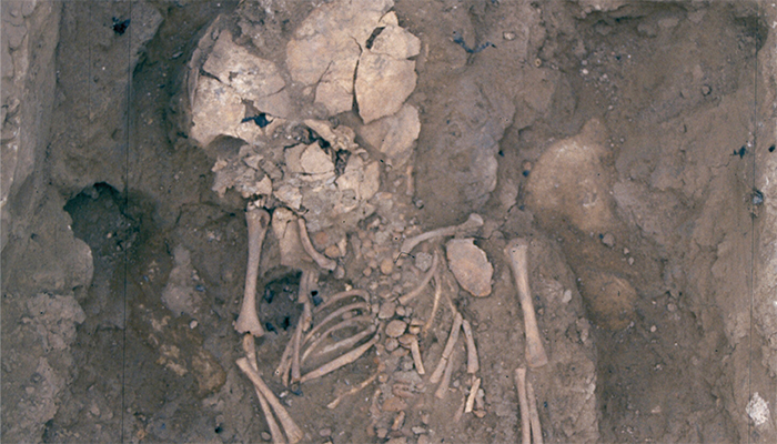
Burial of a perinatal individual from the Fortalesa dels Vilars (Arbeca, Lleida) site.
Could you explain in more detail how the neonatal line in teeth can provide precise information about birth and death timing? Why is this technique more reliable than conventional methods?
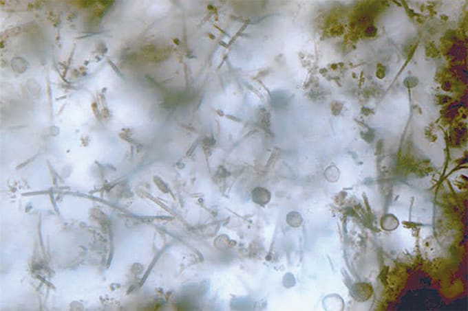
The neonatal line is a unique feature that forms in tooth enamel at birth due to the physiological stress that results from the transition to life outside the womb. It serves as a highly precise marker for determining whether an infant survived birth and, if so, how long they lived afterward. Unlike conventional methods, which estimate age at death based on skeletal development and are often imprecise, the neonatal line allows for the exact chronological determination of age. This is achieved by tracking the formation of enamel both before and after birth.
The key to this precision lies not only in identifying the neonatal line but also in calculating the crown formation time. By measuring the enamel increments (which appear as daily cross-striations) within the tooth, we can determine both the gestational age at the time of birth and how long the infant survived post-birth. The combination of crown formation time and the presence of the neonatal line provides unparalleled accuracy in determining birth and death timing.
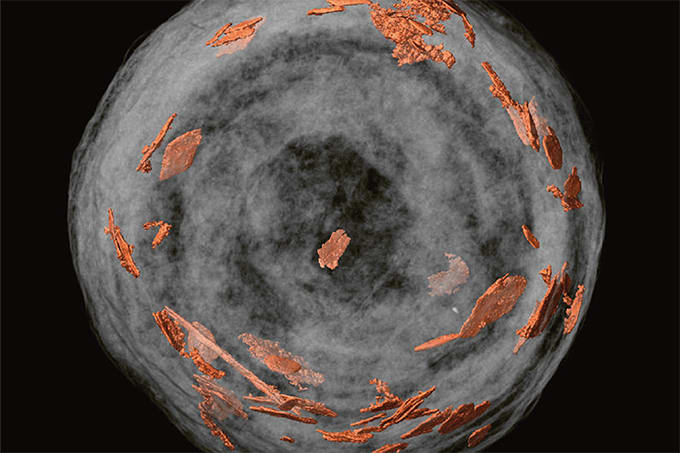
Furthermore, we complement this technique with synchrotron X-ray fluorescence, which detects elemental changes in teeth, particularly fluctuations in zinc and calcium levels, which are closely associated with the birth process.
Any big analytical challenges that you had to overcome?
The most significant challenge we encountered was the fragile nature of tooth germs and their susceptibility to damage from taphonomic processes, which made it difficult to visualize the neonatal line and other enamel growth features. Additionally, it was challenging to distinguish between prenatal and postnatal accentuated lines in archaeological samples, as they can sometimes appear quite similar under specific conditions.
To overcome these challenges, we employed synchrotron X-ray fluorescence to detect elemental changes in the enamel, such as shifts in zinc and calcium levels, which helped confirm the presence of the neonatal line. In addition, we calculated the prenatal crown formation time by measuring the enamel that formed before birth to determine how long the tooth developed in utero. By comparing these measurements with established developmental benchmarks, we could verify whether the line observed was indeed the neonatal line or an accentuated line formed before birth. This dual approach allowed us to confidently confirm the presence of the neonatal line and ensure the accuracy of our findings.
Could the techniques used in this study be applied to other archaeological or anthropological investigations?
Yes, the techniques we developed, particularly the identification of the neonatal line and the use of synchrotron X-ray fluorescence for precise age determination, have broad applications in both archaeology and anthropology. They can be used to reconstruct mortality patterns in different populations and provide valuable insights into maternal and infant health, birth practices, and social structures across various time periods and regions.
Looking ahead, we plan to extend this research to other populations. We are also exploring the possibility of integrating isotopic analysis and synchrotron X-ray fluorescence for additional elements, such as zinc, calcium, and copper, to further investigate demographic trends and environmental influences on health and mortality.


