Coinciding almost exactly with the reign of Henry VIII, the life of the Mary Rose has fascinated historians for years. Having fought many successful battles for the infamous royal, the prized flagship’s career finally came to an end in July 1545, at the Battle of the Solent. It was eventually raised from the seabed in 1982 – along with some 19,000 artefacts – and has since been housed in its own dedicated museum in Portsmouth, UK (maryrose.org).
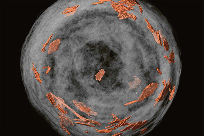
Scientists from the Universities of Warwick and Ghent have now analyzed three artefacts using the XMaS (X-ray Materials Science) beamline facility (1). “Our analysis revealed that the three brass links, believed to be chain mail fragments, were manufactured from an alloy of 73 percent copper and 27 percent zinc,” says Mark Dowsett, lead author of the resulting paper. “This modern alloy composition was consistent across the three samples, indicating that Tudor brass production techniques were very well developed and controlled,” he adds.
The links had also undergone different cleaning and anti-corrosion treatments, enabling the researchers to compare the effectiveness of different conservation efforts. By looking for chemical species that signal corrosion (such as nantokite), the team was able to show that soaking and coating of the artefacts with inhibitors – combined with their storage conditions – had effectively prevented corrosion. “Analysis of this kind can not only inform the overall conservation of the Mary Rose, but also future projects too,” says Eleanor Schofield, Head of Conservation at the Mary Rose.
Interestingly, their analysis also revealed traces of heavy metals embedded in the surface of the artefacts. “Further analysis is needed to confirm their origin, but they could have been picked up from firearm discharge during the 1545 battle, or through extensive WWII bombing of the Portsmouth Dockyard. Otherwise, they might simply indicate contamination caused by rivers flowing into the Solent,” says Dowsett.
The increase in X-ray flux available on the XMaS beamline, combined with its Pilatus camera, facilitated extraordinary sensitivity. “X-ray diffraction doesn’t normally give you a sensitivity of parts per million,” says co-author Mieke Adriaens. “But our unique approach allowed us to merge thousands of camera pixels into each data point in the diffraction pattern, which allowed us to identify low-intensity features that would have otherwise been buried in background noise.”
The overall aim for the research team is to devise new analytical methods using electrochemistry as a means of inducing controlled chemical modifications that can be studied in real time using techniques such as synchrotron X-ray diffraction or X-ray absorption spectroscopy.
Currently, the XMaS beamline is being rebuilt in a bid to reduce beam size and increase the X-ray energy range. “This is very exciting for us – we hope to win beam time on the new instrument for further work on other Mary Rose samples,” says Dowsett.
References
- MG Dowsett et al., J Synchrotron Rad (2020) 27, 653-663. DOI: 10.1107/S1600577520001812


