Conexiant
Login
  • The Analytical Scientist
  • The Cannabis Scientist
  • The Medicine Maker
  • The Ophthalmologist
  • The Pathologist
  • The Traditional Scientist
The Analytical Scientist
  • Explore

    Explore

    • Latest
    • News & Research
    • Trends & Challenges
    • Keynote Interviews
    • Opinion & Personal Narratives
    • Product Profiles
    • App Notes

    Featured Topics

    • Mass Spectrometry
    • Chromatography
    • Spectroscopy

    Issues

    • Latest Issue
    • Archive
  • Topics

    Techniques & Tools

    • Mass Spectrometry
    • Chromatography
    • Spectroscopy
    • Microscopy
    • Sensors
    • Data & AI

    • View All Topics

    Applications & Fields

    • Clinical
    • Environmental
    • Food, Beverage & Agriculture
    • Pharma & Biopharma
    • Omics
    • Forensics
  • People & Profiles

    People & Profiles

    • Power List
    • Voices in the Community
    • Sitting Down With
    • Authors & Contributors
  • Business & Education

    Business & Education

    • Innovation
    • Business & Entrepreneurship
    • Career Pathways
  • Events
    • Live Events
    • Webinars
  • Multimedia
    • Video
Subscribe
Subscribe

False

The Analytical Scientist / Issues / 2013 / Feb / Camera Focus
Mass Spectrometry

Camera Focus

How mass spectrometric imaging of complex surfaces, such as animal tissue, is being expanded with a position- and time-sensitive pixelated detector

By Ron M. A. Heeren, Julia H. Jungmann 02/20/2013 1 min read

Share

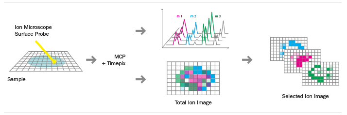
In high-throughput time-of-flight mass spectrometric imaging (TOF-MSI), the pressure is on the detection system: it must measure the impact position and time of the entire ion load from an ionization event in a single measurement frame. Now, using an innovative electron and ion imaging camera, this can be achieved. The approach combines a chevron microchannel plate (MCP) stack with an active pixel detector chip. Ion mass microscopy is a molecular imaging technique that delivers analyte identity and spatial localization with very high throughput at sub-micron pixel sizes. A large-area desorption/ionization beam illuminates the sample surface; ion optics magnify the molecular images and retain the spatial information; and the molecular ion distributions are mapped on a position-sensitive detector.

Camera Focus Graphic

The new detection system is based on application-specific integrated circuits (ASIC) developed by the Medipix collaboration hosted by CERN, Geneva, Switzerland. The Timepix chips comprise tens of thousands of pixels each with a pitch of 55 µm, all of which function as individual units for parallel detection. Importantly, the Timepix chips provide spatial event information (via the pixel address) as well as time-of-flight, particle energy and event counting at the pixel level.

The new camera has been successfully tested on benchmark systems (protein and lipid standards) and with biologically relevant macromolecular tissue samples (rat brain and testes). TOF-MSI systems offer several unique capabilities afforded by the combination of high signal-to-noise ratios, multiplexed detection of events by a highly parallel detection system, high sensitivity, dynamic range, large mass range and simultaneous detection of position- and time-information by a single detector. All of which means higher throughput for faster analysis, the ability to detect small quantities of analytes, and better spatial resolution, revealing unprecedented detail.

As TOF-MSI continues to mature, applications in diagnostics, medical imaging, fundamental atomic and molecular physics, and even space science are likely to grow.

For more information, visit www.amolf.nl/medipix.

Newsletters

Receive the latest analytical science news, personalities, education, and career development – weekly to your inbox.

Newsletter Signup Image

About the Author(s)

Ron M. A. Heeren

Director, M4I, and Distinguished Professor, University of Maastricht, Netherlands

More Articles by Ron M. A. Heeren

Julia H. Jungmann

Julia Jungmann obtained her MSc degree in Experimental Physics from the University of Groningen, The Netherlands. In 2007, she joined the instrumentation group at the Kernfysisch Versneller Instituut in Groningen for a feasibility study for detecting the amount of gamma-emitting radionuclides suspended in water with advanced algorithms for real-time signal processing. In 2012, Julia obtained her PhD at the University of Utrecht. “My doctoral project focused on the implementation and application of state-of-the art pixelated solid state detectors to biomolecular mass spectrometry imaging carried out in the Biomolecular Imaging Mass Spectrometry group of Professor Ron M. A. Heeren at the FOM-Institute AMOLF in Amsterdam.” Her research interests include imaging and detections systems as well as intelligent sensor systems for biomolecular, biomedical and environmental applications.

More Articles by Julia H. Jungmann

False

Advertisement

Recommended

False

False

The Analytical Scientist
Subscribe

About

  • About Us
  • Work at Conexiant Europe
  • Terms and Conditions
  • Privacy Policy
  • Advertise With Us
  • Contact Us

Copyright © 2025 Texere Publishing Limited (trading as Conexiant), with registered number 08113419 whose registered office is at Booths No. 1, Booths Park, Chelford Road, Knutsford, England, WA16 8GS.