Conexiant
Login
  • The Analytical Scientist
  • The Cannabis Scientist
  • The Medicine Maker
  • The Ophthalmologist
  • The Pathologist
  • The Traditional Scientist
The Analytical Scientist
  • Explore

    Explore

    • Latest
    • News & Research
    • Trends & Challenges
    • Keynote Interviews
    • Opinion & Personal Narratives
    • Product Profiles
    • App Notes

    Featured Topics

    • Mass Spectrometry
    • Chromatography
    • Spectroscopy

    Issues

    • Latest Issue
    • Archive
  • Topics

    Techniques & Tools

    • Mass Spectrometry
    • Chromatography
    • Spectroscopy
    • Microscopy
    • Sensors
    • Data and AI

    • View All Topics

    Applications & Fields

    • Clinical
    • Environmental
    • Food, Beverage & Agriculture
    • Pharma and Biopharma
    • Omics
    • Forensics
  • People & Profiles

    People & Profiles

    • Power List
    • Voices in the Community
    • Sitting Down With
    • Authors & Contributors
  • Business & Education

    Business & Education

    • Innovation
    • Business & Entrepreneurship
    • Career Pathways
  • Events
    • Live Events
    • Webinars
  • Multimedia
    • Video
Subscribe
Subscribe

False

The Analytical Scientist / Issues / 2024 / Oct / 3D Atlas of the Developing Brain
Spectroscopy Microscopy Translational Science

3D Atlas of the Developing Brain

Researchers combine MRI and light sheet fluorescence microscopy to create a dynamic framework for studying brain development and neurodevelopmental disorders

10/31/2024 1 min read

Share

Credit: Kim Lab / Penn State Creative Commons

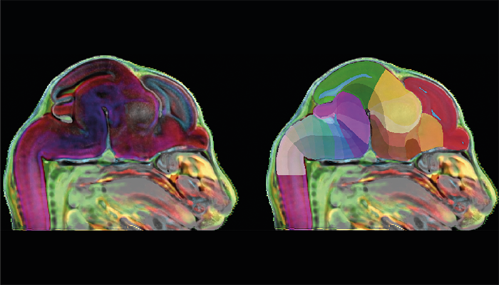
A 3D atlas of the developing mouse brain has been created by researchers at Penn State College of Medicine using high-resolution imaging techniques. The atlas represents a common reference framework for analyzing brain architecture in both normal and diseased states, and it could be used to study brain development as well as neurodevelopmental disorders.

"Maps are a fundamental infrastructure to build knowledge upon, but we don't have a high-resolution 3D atlas of the developing brain," said Yongsoo Kim, associate professor of neural and behavioral sciences at Penn State College of Medicine and senior author on the study, in a press release. "We are generating high-resolution maps that we can use to understand how the brain grows under normal circumstances and what happens when a brain disorder emerges."

The team employed magnetic resonance imaging (MRI) to capture the overall form and structure of the brain across seven developmental time points – from the embryonic period to the immediate postnatal stage. The researchers also used light sheet fluorescence microscopy (LSFM) to visualize the developing brain at single-cell resolution, enabling precise mapping of brain cells and structures. By integrating these two imaging techniques, the researchers created a unified 3D framework – the Developmental Common Coordinate Framework (DevCCF).

"Without this 3D map of the developing brain, we cannot integrate data from emerging 3D studies into a standard spatial framework or analyze the data in a consistent manner," Kim added.

Putting the atlas into practice, the team investigated GABAergic neurons, a type of nerve cell involved in crucial brain functions and implicated in several neurological disorders, including schizophrenia and autism. The researchers were able to trace the development of GABAergic neurons across different brain regions, providing insights into how these cells arise during normal development – and what might go wrong in disease states.

The 3D atlas is freely accessible through an interactive web-based platform and, according to Kim, “It will drive the next evolution of brain research driven by machine learning and artificial intelligence." 

Newsletters

Receive the latest analytical science news, personalities, education, and career development – weekly to your inbox.

Newsletter Signup Image

False

Advertisement

Recommended

False

Related Content

The Analytical Scientist Innovation Awards 2024: #3
Spectroscopy
The Analytical Scientist Innovation Awards 2024: #3

December 6, 2024

4 min read

Bruker’s multiphoton microscopy module, OptoVolt, ranks third in our Innovation Awards. Here, Jimmy Fong, product development lead, walks us through the major moments during development.

More Bang for Your Buck
Spectroscopy
More Bang for Your Buck

December 4, 2024

1 min read

Researchers develop more stable catalysts for dry reforming of methane – a promising method for carbon capture and utilization (CCU)

The Analytical Scientist Innovation Awards 2024: #1
Spectroscopy
The Analytical Scientist Innovation Awards 2024: #1

December 10, 2024

2 min read

And the technology ranked first in our 2024 Innovation Awards is…

The Analytical Scientist Innovation Awards 2024
Spectroscopy
The Analytical Scientist Innovation Awards 2024

December 11, 2024

10 min read

Meet the products – and the experts – defining analytical innovation in 2024

False

The Analytical Scientist
Subscribe

About

  • About Us
  • Work at Conexiant Europe
  • Terms and Conditions
  • Privacy Policy
  • Advertise With Us
  • Contact Us

Copyright © 2025 Texere Publishing Limited (trading as Conexiant), with registered number 08113419 whose registered office is at Booths No. 1, Booths Park, Chelford Road, Knutsford, England, WA16 8GS.