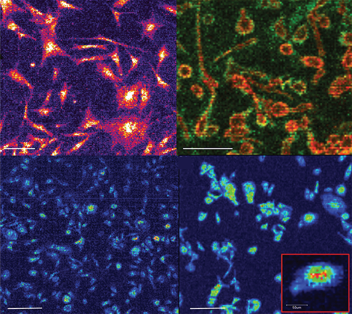
Molecular images of single cells in 2D cultures generated on a MALDI-ToF instrument. Top: fibroblasts (left) and macrophages (right); bottom: cardiomyocytes (left) and breast cancer cells (right). Inset: a zoom in on a single breast cancer cell showing the different levels (blue, green, red) of phosphatidylcholine 36:1 in the cell. Scale bars are 200 μm; inset scale bar is 50 μm.
Eva Cuypers will be featured in our upcoming series of articles on mass spec imaging for cancer. So keep your eyes peeled in the New Year!


