When were the dyes used by artists and craftsmen for pigments and painting introduced, what technology did this require, and what do these insights shed on the times?
- A leather fragment of a quiver (see figure 1), found in an archaeological excavation led by the Metropolitan Museum in Egypt in 1911-1912. The fragment, which measures 11cm by 13cm, dates to the Middle Kingdom, ca. 2124-1981 BC, and was found in a tomb in Thebes, el-Khokha, Upper Egypt.
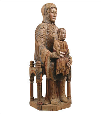
- A small fragment from the purple robe of the Child in a French 12th century sculpture called Virgin and Child in Majesty. The sculpture (see figure 2), stands 79.5cm high and is from the Auvergne, France It was a gift of J. Pierpont Morgan in 1916.
- To detect compounds that make up a very small percentage of the final product.
- To use minimally invasive techniques.
For millennia, artists and craftsmen have used colored organic compounds to dye fabrics and to create pigments for use in painting. These dyes, such as madder dye, cochineal, logwood, and many others, were extracted from plants and insects. Quite naturally, the most successful dyes were those with the highest tinting strength. A clear advantage for a dyer, the need to purchase only small amounts of dye to tint several pounds of yarn is the bane of the analytical chemist: detecting compounds that make up a very small percentage of the final product. To make things more difficult, the analysis of works of art should ideally be conducted with non-destructive techniques, and when sampling is necessary it should be limited to almost invisible amounts. In the case of dyes, non-invasive methods are of little help. UV-Vis reflectance spectra lack sufficient specificity, there is no X-ray fluorescence elemental signature characteristic of each dye, and most dyes are highly fluorescent under laser excitation, essentially hiding their Raman spectrum.
SERRS is not affected by the above limitations. Natural and synthetic organic colorants are, in general, aromatic or heterocyclic compounds (or, in the case of the carotenoids, highly conjugated linear molecules), and when adsorbed on the surface of metallic nanoparticles they experience an enormous enhancement in Raman scattering. Analysis of works of art has become one of the most significant applications of the technique, which is routinely applied to the identification of organic colorants at the Metropolitan Museum of Art. The high sensitivity of SERRS makes it possible to identify organic colorants in textiles, drawings, paintings, and other polychrome objects, from samples as small as 25 micrometers in diameter.
Samples from art, as well as being microscopic, are invariably very complex. On top of that, the objects we routinely analyze are extremely old: aging and deterioration transform the target molecules and produce interfering species. Yet, SERRS has proven to be surprisingly immune to such interference. Part of the success lies in the microanalytical protocol we developed after lengthy trials. We work at resonant excitation, using a monodisperse silver colloid produced by microwave-supported reduction of silver sulphate with glucose and sodium citrate, and we often employ a two-step measurement approach: analysis of the sample as-is, followed by recovery of the sample and a second analysis after a lossless non-extractive hydrolysis sample treatment. All work is performed with a Bruker Senterra Raman spectrometer, using 488nm laser excitation. We have successfully demonstrated that the colloid we use is stable over long periods; that the sample treatment protocol enhances sensitivity over comparable approaches, and that the spectra obtained can be cross-referenced with digital spectral libraries using common software.
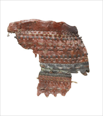
Using SERRS we identified alizarin, a molecule obtained from the root of the madder plant (Rubia sp.), in a microscopic sample from the red tinted surface of the leather quiver. This is so far the oldest scientifically documented evidence for the use of natural dyes. Madder dye is one of the most stable red dyes and was commonly used from antiquity to the late 19th century, not only as a mordant dye for fabrics, but also as a pigment (properly a lake, a complex of the molecule with an inorganic cation such as aluminium, calcium, or tin). The ancient Egyptians had mastered the art and science of extracting the dye from the root (the process requires alkali at low temperatures) and precipitating it as a pigment using alum under carefully controlled pH and temperature. This was 4,000 years ago. Older examples of such knowledge surely exist in other museums: with SERRS, we now have a way to find out how far dye chemistry goes back in time – with essentially no impact on the precious archaeological evidence.
The analysis of the purple robe of the Child showed that lac dye, a colorant extracted from an insect native of South East Asia had been used. This is the first evidence for this colorant in Europe: the fact that it reached southern France from India at the time of the crusades points to the extent and strength of global trade networks. These results – the foundation of our work on the analysis of art – can be translated to other fields, making SERRS more easily applicable in food and cosmetics studies, environmental analysis, and forensic science.
Marco Leona is the David H. Koch Scientist in Charge at the Department of Scientific Research, The Metropolitan Museum of Art, New York, USA.
Please read the other articles in this series:
Casting Light on Renaissance Illuminations
Deconvoluting the Creative Process
Understanding Ancient Prescriptions
Newsletters
Receive the latest analytical science news, personalities, education, and career development – weekly to your inbox.


