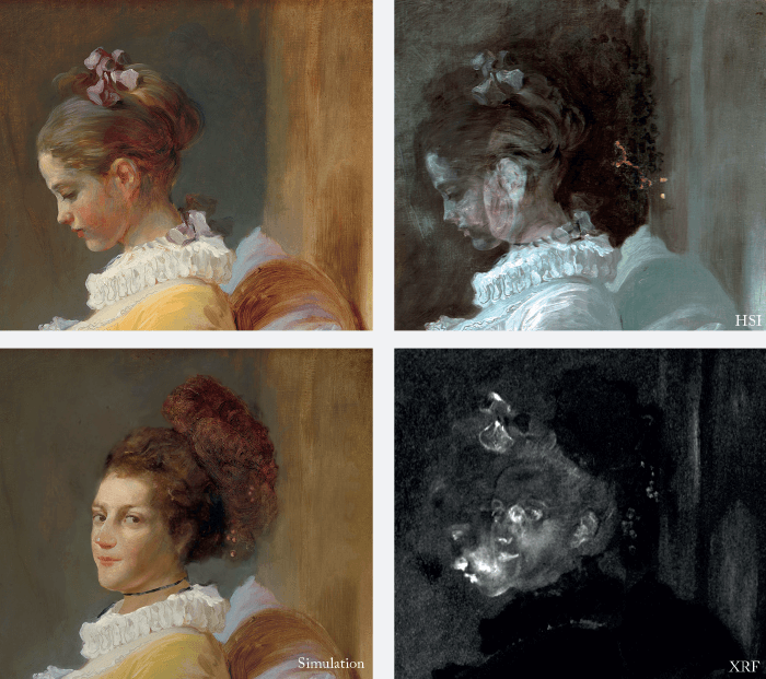
The paintings of Jean-Honoré Fragonard (1732–1806) are known for their brilliant coloring and alla prima brushwork. The National Gallery of Art’s Young Girl Reading is one of the most beloved examples of his virtuoso painterly style. In 2012, the surprise rediscovery of a drawing by Fragonard, further confirmed the rarity of Young Girl Reading. This remarkable sheet is covered with “thumbnail-sized” sketches, each showing a single figure. These sketches clearly relate to Fragonard’s so-called “fantasy figures,” a series of boldly-colored paintings of individual models in extravagant fancy dress. Today, the “fantasy figures” are distributed amongst the most distinguished public and private art collections in the world, including the Musée du Louvre and the Metropolitan Museum of Art.
At the Gallery, the revelations of the drawing prompted a two-year investigation of Young Girl Reading, conducted as a collaborative effort by the authors. Our findings indicated that underneath the profile of Young Girl Reading lay another face entirely, that of a woman turned outwards, looking directly at the viewer. This now-hidden woman once had a large plumed and beaded headdress, similar to those of the “fantasy figures.” Scientific standoff chemical imaging (RIS, XRF and RTI) and study of a cross-section taken in an area of change indicated that the artist first intended to depict a woman looking outwards, holding a book. He completed this composition, then left it alone, perhaps for as long as a year before returning to adjust the orientation of the head of his model, repurposing it as the Young Girl Reading known today. In her original pose, Fragonard’s woman holding a book corresponds to one of the sketches on the newly-discovered drawing, thereby confirming that the painting underlying Young Girl Reading was conceived as a “fantasy figure.” Using similar multidisciplinary methods (art-historical research, conservation science, imaging techniques), the authors are currently researching other “fantasy figures” in the hopes of shedding further light upon Fragonard’s conception of and approach to the ensemble as a whole. Our findings will be presented with the forthcoming exhibition, Fragonard’s Fantasy Figures, at the National Gallery of Art, Washington, from October 8 through December 3, 2017.
The Toolbox
Multi-modal imaging – diffuse reflectance imaging spectroscopy (RIS), x-ray fluorescence (XRF) imaging, reflectance transformation imaging (RTI) and high-resolution color photography – were used to reveal as much as possible about the underlying painting beneath Young Girl Reading. Diffuse reflectance imaging from 1000 to 2500 nm was performed with a recently designed and constructed hyperspectral near-infrared imaging camera with 2.7 nm spectral sampling. Narrow spectral band NIR reflectance imaging has been found to give some of the clearest images of underlying paint compositions or underdrawing, as well as aiding in the mapping of artist materials (pigments & paint binders). XRF imaging spectroscopy, using a home-built scanning sensor, provided elemental maps to help complete the ‘picture’ of the underlying figure (particularly useful were maps for lead, mercury and iron). John K. Delaney is senior imaging scientist, Yuriko Jackall is specialist in eighteenth-century French paintings, and Michael Swicklik is senior conservator of paintings, all at the National Gallery of Art, Washington, USA.Analytical palette
It’s fair to say we rely pretty heavily on X-rays! X-ray fluorescence (XRF), for example, allows us to discover the elemental content of materials. Handheld XRF systems are now used throughout the world by anyone who has access to basic funding, which has really opened up this kind of research. Nowadays, they have essentially become black box instruments. Basically, you irradiate a square centimeter of a painting and figure out – indirectly – which pigments were used to paint the different colored areas. The high-end of the X-ray analysis spectrum relies on synchrotron radiation-based nano and microbeams (aka as synchrotron X-ray microprobes), which can be used to analyze tiny samples (a few 100 micrometers across) from famous paintings to investigate natural degradation processes. Depending on which pigments have been used and in what combinations, colors can change as they interact with physical or chemical agents in the environment. For instance, some of the reds that have been used by Rubens develop a degradative black or a white crust. Degradations such as these follow a sequence of chemical modifications and we can find traces of these sequences if we analyze paint samples at a very high spatial resolution. For example, vermilion red – mercury sulfide – transforms gradually to mercurous chloride (calomel) over three or four steps, involving exotic compounds called mercury sulfochlorides; our advanced imaging techniques allow us to see the difference between all these compounds and therefore to trace back the gradual transformation process from the sulfide to the chloride. And electrochemical methods help support our work in this area by assessing potential routes of degradation ahead of time. Degradation information is important to conservators, who can investigate preventative conservation to shield or isolate the works of art from chemical agents that are likely to have a negative impact. Another activity that is more directly aimed towards art historians and museum people moves away from the nano- or microscopic level, involves developing instruments that allow chemical imaging at the square meter scale. Such macro-scanning X-ray fluorescence (MA-XRF) instruments provide large-scale chemical maps that show both the painting and the distribution of chemical elements within it. We cannot only show what’s on the surface, which allows art historians to learn about the materials the artist has used, but also what is present below the visual surface. If an artist has, during the creative process, changed his mind and overpainted part of the painting with something else (known as ‘pentimento’ in art circles), our X-ray “eyes” will see it. Such information can be highly relevant for art historians who seek to reconstruct the history of or understand the creative process behind a work of art.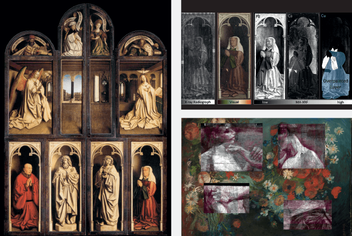
Chefs d’oeuvre
The 15th century Ghent Altarpiece (also called the Adoration of the Mystic Lamb or The Lamb of God) is currently housed in Saint Bavo Cathedral, Ghentand is considered one of the van Eyck brothers’ masterworks. Using our MA-XRF scanner, we have been investigating a number of the panels over the last few years to help restorers, who suspected that the polyptych, as it is today, is not entirely original. Rather, the Ghent Altarpiece is actually an overpainted version of the original, which is still underneath; only the original was painted by the van Eycks. Despite their strong suspicions, the restorers could not produce compelling enough evidence to convince their colleagues that the upper layers should be removed to reveal again the van Eyck original – until we came along. In such a major restoration, you not only have the restorers, who are the executive force, but also one or more international advisory committees. The restorers aimed to convince all stakeholders that such a painting should be restored to principles observed today, which means keeping original paint and removing all more recent overpaints. So we helped them by scanning the panels and finding visual ways of making the distinction between original van Eyck paint and the paint that was added ca 100 years later. Translating the very strong hunch of the conservation team into images meant that their international colleagues could be persuaded to remove all overpaint. The consequence? The removal of around 60 percent of the total paint! Clearly, this is invasive, but the original van Eyck paint will become visible again after being covered for around 400 years. I’m sure it will have a big impact and lasting consequences.For another example, I’ll skip from the 15th century to the 19th century. We have studied quite a few paintings by Vincent van Gogh over the last ten years (and, in fact, the first high-level painting we analyzed with our new imaging method was also by van Gogh). However, this example involves a specific piece associated with my hometown of Antwerp, which he painted while he was at the academy here. Van Gogh has a Dutch period, a short Antwerp period of only one month, and then he moved to Paris, where he met the Impressionists and continued on to Provence. While in Paris, he encountered a completely new way of painting and came to dislike a number of his previous darker and less colorful works. He decided to paint over them, which is why there are a number of double-layered van Gogh paintings. We’ve examined a number of these, including Still Life with Meadow Flowers and Roses – a painting that is now in the Kröller-Müller Museum in the north of Holland. It was originally bought as a van Gogh but, at some point in the 1990s, was rejected as the real deal. Why? Because it was the wrong size, for one thing. Indeed, the composition of the surface painting was not typically van Goghian, according to the art experts. It was removed from the catalogue of van Gogh paintings and was an orphan for about 20 years. In 2012, we reanalyzed the work with MA-XRF and showed, much more clearly – brushstrokes and all – the image of two wrestlers grasping each other by the arms beneath the surface. By combining that clearer image with what was understood of van Gogh’s work in Antwerp, we could strengthen the link between the painting and van Gogh. As a result, the art historians of the van Gogh museum in Amsterdam and the Kröller-Müller museum reinstated the painting as a van Gogh the same year. Koen Janssens is Full Professor, general and analytical chemistry, and Vice-Dean, Faculty of Sciences, at the University of Antwerp, Belgium.
The late paintings of Rembrandt van Rijn pose significant challenges because of their multi-layer build up, heterogeneous materials, compositional changes, degradation problems and complex histories. In The Sampling Officials of the Amsterdam Drapers’ Guild from 1662, better known as The Syndics (Rijksmuseum Amsterdam), Rembrandt radically altered the position of several of the figures. Developments in analytical science are now making it possible to start answering some of the questions that arise. In 2013, the painting was investigated with portable macro-X-ray fluorescence scanning (MA-XRF) in the galleries of the Rijksmuseum using the Bruker M6 scanner. A series of 15 overlapping scans were captured over a period of a few days. The resulting elemental distribution map of lead associated with the pigment lead white, provides additional information regarding the repositioning, some four times, of the figure of the steward in the background, Frans Hendricksz Bel, which appears to have been fully painted in his former locations, at the far right and in between the two sampling officials – before being placed behind the two syndics in the centre of the composition. Elemental distribution maps for cobalt, nickel, arsenic and potassium associated with the pigment smalt also suggest that the hat of Jochem de Neve, the syndic to the right of Bel, was also reduced to accommodate the final position of Bel, although this change could also relate to the ranking of the officials. This offers new information on the much debated order, function and meaning of the changes. The X-radiograph and infrared reflectography (IRR) had already provided some information but were difficult to interpret. It is more often the case that traditional imaging techniques, such as X-radiography and IRR, prove ineffective in the study of late Rembrandt paintings due to Rembrandt’s choice of materials, particularly his use of radio-transparent quartz and earth-rich grounds, and pigments, such as organic lakes, smalt, earths and bone black.
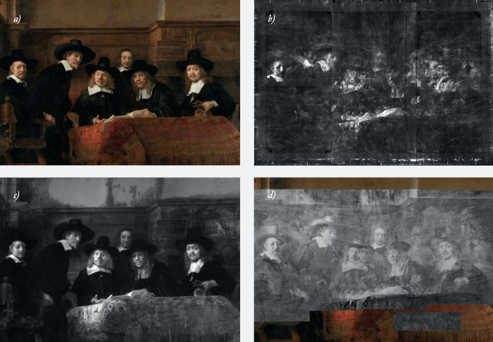
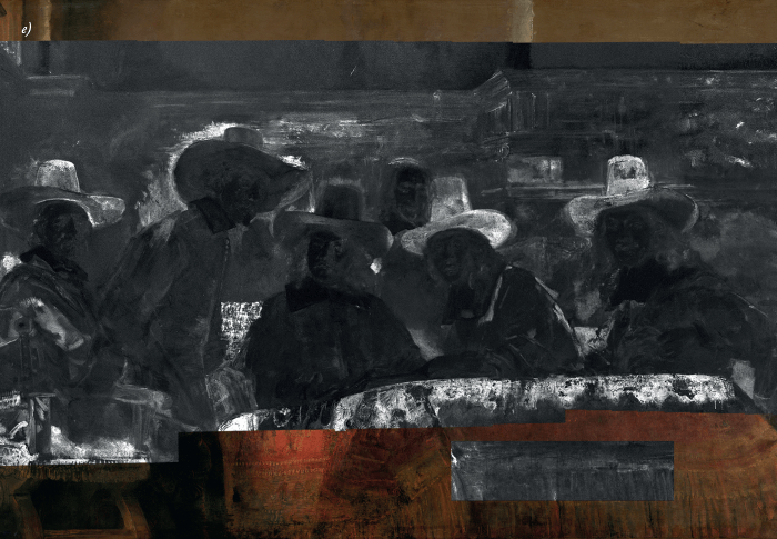
In this project, scientists, conservators and art historians are working together to develop and apply new imaging techniques to the study of (late) Rembrandt paintings. Partners include: Joris Dik (Delft University of Technology), Koen Janssens (University of Antwerp) and John Delaney (The National Gallery of Art, Washington) as well as: Mauritshuis, The Hague, Netherlands Institute for Art History (RKD), Cultural Heritage Agency of Netherlands (RCE), the Wallraf-Richartz Museum, Cologne and Synchrotron Soleil France. The MA-XRF scanning of the painting was carried out by Geert van der Snickt (University of Antwerp), Anna Krekeler and Lisette Vos. Results of this particular painting were presented at the Painting Techniques, History, Materials and Studio Practice, 5th International Symposium, (Rijksmuseum, 18-19-20 September 2013) and are illustrated in J. Bikker and A. Krekeler, “Experimental technique: The Paintings”, in J. Bikker et al., [exh. cat], Rembrandt: The Late Works, 2014. Petria Noble is Head of Paintings Conservation at the Rijksmuseum and is researcher in the Science4Arts- ReVisualising late Rembrandt project (2012-2018) sponsored by the Netherlands Organization for Scientific Research (NWO) and the National Science Foundation.
There is more than meets the eye in The Portrait (1935), a painting by renowned Belgian artist René Magritte. In the course of examining and treating the painting for an upcoming exhibit at MoMA in 2014 entitled Magritte: The Mystery of the Ordinary, 1926-1938, the museum conservators took an X-ray image of the painting and discovered an underlying composition representing the head and torso of a female nude (2; see Image 2). In fact, the hidden image corresponds to the upper left section of The Enchanted Pose, also painted by Magritte in 1927, and presumed lost since the early 1930s. The lower half of the nude was discovered shortly after under The Red Model (1935) and the quest continues for the remaining missing half of the original painting. Magritte painted right over the section of the painting, but the underlying paint in still visible around the edges of the painting and around the edges of the bottle. He also incorporated passages of the underlying composition in the new one, for example, in the brown reflection in the glass. Why Magritte chose to sacrifice this composition and reuse the canvas remains a mystery. Maybe it was out of resentment after The Enchanted Pose was rejected for a 1932 group show in Paris, or he may have simply run out of canvas, or perhaps the work got damaged and could no longer be exhibited. Even if Magritte did not intend for us to see this painting, this study has allowed us to virtually reconstruct an approximate visual record of it and, in the process, help us understand his artistic development by studying what he created – but also what he chose to obliterate.
The toolbox
To learn more about the materials Magritte used for both top and underlying compositions, the painting was imaged using macroscopic X-ray fluorescence analysis (MA-XRF; Bruker), which maps the distribution of chemical elements that are representative of the pigments in the paints. The face of the hidden figure is revealed in both the lead and iron maps, suggesting it was executed with earth pigments and lead white-based paints. The mercury map, on the other hand, shows the lips were retouched with a paint containing vermillion. The sky around the figure is visible in the chromium map indicating the paint contains chromium oxide green, a bluish green pigment. The same paint appears in other late 1920s paintings by Magritte and has a similar composition as a blue household paint manufactured by Ripolin (3). The bottle in the upper composition is visible in the calcium and cadmium map, suggesting Mondrian modified the tonality of the bone black-based paint by mixing with cadmium yellow paint.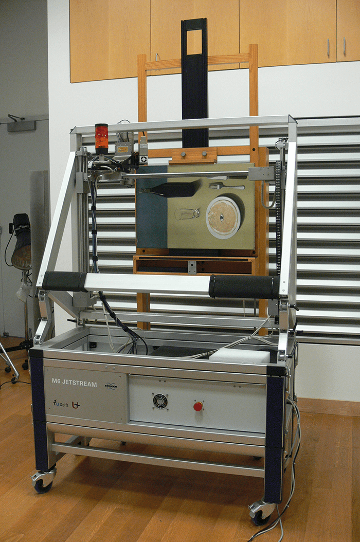
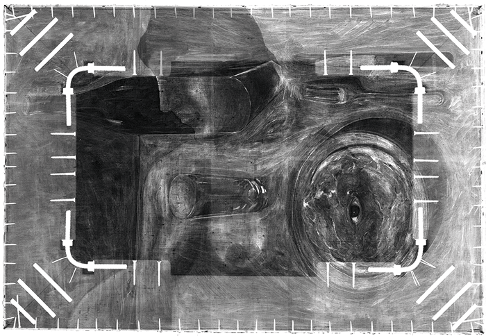
In total, twelve chemical elements were identified by MA-XRF and the stratigraphy of the painting (see Figure 2) can be inferred from the corresponding distribution maps and some complimentary analysis (4). This technique provided valuable insight into both the creative process and choice of materials of this artist in a noninvasive way, without the need of taking cross sections. Ana Martins is a research scientist in the conservation department of the Museum of Modern Art (MoMA), New York, USA.
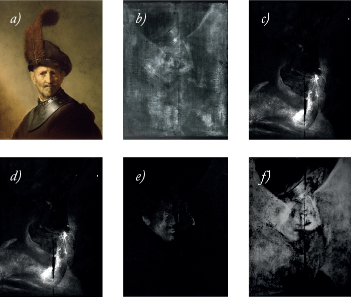
An Old Man in Military Costume, painted by Rembrandt in 1630-31, is not only a compelling portrait of an old man wearing a cloak, a neck protector (known as a gorget) and a feathered cap, it is also an analytical challenge: beneath the painting of the old man lies a second, hidden painting. How can analytical science reveal the underlying image without causing any damage to the upper painting? The answer is X-rays (and gamma rays) – and patience. In 1978, X-radiography first revealed the face of a man, rotated 180 degrees from the upper portrait. But who was he, how was he dressed, and what colors were used? Patience. In the early 1990s, neutron activation autoradiography (NAAR) was carried out and, although many of the NAAR images contained contributions from multiple elements, some tantalizing information was revealed: mercury (from the pigment vermilion, HgS) was used to paint the face, and he was wearing a cloak painted with a copper-containing pigment. But the details were fuzzy. More patience. In 2013, the painting was studied by macro-X-ray fluorescence (MA-XRF) scanning, which enabled complete distribution maps of individual elements to be collected. The results allowed us to create a digital reconstruction of the underlying painting: a young man, wearing a cloak with a wide collar – perhaps also a gorget. But what, if anything, is he wearing on his head? Nothing is evident in the XRF maps, but they cannot detect the lower Z elements that comprise organic materials. Even more patience... Analytical science will, someday, provide us with a tool to answer this and other questions.
Collaboration
To gain a better understanding of the materials and methods used by one of the world’s greatest artists, this, as with most cultural heritage research, was a collaboration between scientists, conservators and art historians: Karen Trentelman, senior scientist and leader of the Technical Studies research group at the Getty Conservation Institute; Koen Janssens and Geert van der Snickt, X-ray Analysis, Electrochemistry and Speciation, University of Antwerp; Joris Dik, Laboratory of Materials Science, Delft University of Technology; Yvonne Szafran, head of Paintings Conservation at the J. Paul Getty Museum; and Anne Woollett, curator of paintings at the J. Paul Getty Museum.Newsletters
Receive the latest analytical science news, personalities, education, and career development – weekly to your inbox.

References
- Yuriko Jackall, John K. Delaney, and Michael Swicklik, “‘Portrait of a woman with a book’: a ‘newly discovered fantasy figure’ by Fragonard at the National Gallery of Art, Washington.” The Burlington Magazine, CLVII (April 2015): 248-254. M Duffy and A Albertson, “The Discovery of Magritte’s ‘The Enchanted Pose’, Inside/Out, a MoMA-PS1 Blog, The Museum of Modern Art, New York, NY. http://www.moma.org/explore/inside_out/2013/10/31/the-discovery-of-magrittes-the-enchanted-pose. G Gautier et al, “Chemical Fingerprinting of Ready-Mixed House Paints of Relevance to Artistic Production in the First Half of the Twentieth Century. Part I: Inorganic and Organic Pigments”, Appl Spectrosc 63, 597-603 (2009). G Van der Snickt G et al. “Exploring a hidden painting below the surface of René Magritte’s ‘Le portrait’.” Le Portrait. Appl Spectrosc, 70, 57–67 (2016).


