Sodium channels are key in the establishment of action potentials (the drivers of our heartbeat), and dysfunction of voltage-gated sodium channel 1.5 (Nav1.5) in the heart can trigger life-threatening arrhythmias. Despite their significance, however, little is known about the structure and physiological function of these channels.
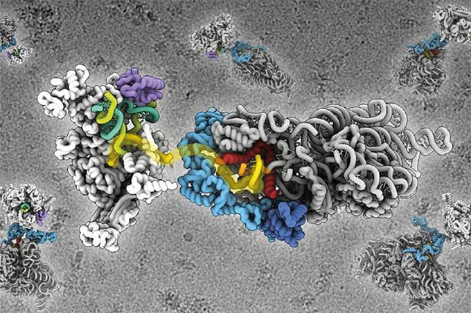
William Catterall and colleagues used cryo-electron microscopy to construct a complete model of Nav1.5’s structure at a resolution of 3.5 Å (1). The result? A more comprehensive understanding of what makes the channels unique – namely, an unexpected glycosyl moiety and the loss of disulfide bonding capability at specific subsites – and advanced insight into mechanisms of voltage-dependent channel functions and sodium ion conduction. The team was keen to understand how antiarrhythmic drugs were interacting with Nav1.5 at an atomic level. “We’ve been able to characterize how flecainide – a commonly prescribed antiarrhythmic – specifically targets the central pore cavity of Nav1.5, physically blocking sodium permeation,” says Catterall.
Looking ahead, the team plan to image multiple arrhythmic mutations and antiarrhythmic drugs at the atomic level. “This will provide much-needed chemical information, which will facilitate structure-based drug design.”

References
- D Jiang et al., “Structure of the cardiac sodium channel”, Cell, 1, 122 [Epub ahead of print] (2020). DOI: 10.1016/j.cell.2019.11.041


