The science department at the Rijksmuseum encompasses a large group of scientists with different specialisms. We conduct research on our collection in close collaboration with conservators, curators and (technical) art historians, with the aim of better understanding, managing and presenting the collection. At HIMS, we study fundamental chemical processes in paints. Dividing my time between the two roles means that I can function as a bridge (or translator) between the art and academic fields.
Fading beauty
We work with conservators to solve the diverse problems they face in conserving and restoring traditional paintings. It is extremely important that research on paintings is carried out before a conservation treatment starts to be able to select the most suitable conservation treatment to preserve and present the painting.
I specialize in aging and degradation studies of pigments and oil paintings at the microscopic and molecular level, especially the interaction between pigment and binding medium. An example of a degradation phenomenon we investigate in detail is the formation of metal soaps in oil paintings, the result of a reaction between the lead or zinc pigment and the oil binder. This phenomenon was first observed in 1997 during the restoration of Rembrandt’s “Anatomy lesson of dr. Nicolaes Tulp” (Mauritshuis, The Hague) and since then has been widely investigated in traditional and modern paintings.
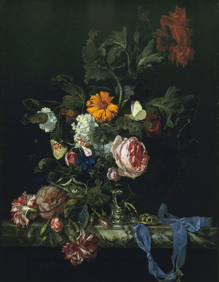
The impact of pigment degradation is clearly visible in the fading of the yellow orpiment paint in “Still Life with Flowers and a Watch” by Abraham Mignon (ca. 1660–1679) (pictured). The yellow and orange arsenic sulfide pigments in the Rosa rubiginosa have degraded after exposure of light, resulting in loss of the flower leaves. We are studying the pathways of the degradation, migration processes of the degradation products and the conditions under which new complexes are formed in the paint.
The future is bright
On a daily basis in my lab, I use mainly imaging-attenuated total reflection (ATR)-FTIR, SEM-EDX, py-GC-MS and Raman. However, I frequently make use of synchrotron facilities to study low-concentration materials at high resolution. The paint sample we are investigate are around 300×100×100 microns in size. Within these samples we study pigment, binders and degradation materials at sub-micron to nanoscale. Synchrotron techniques we have used include XANES, EXAS, SAXS, micro-X-ray diffraction (XRD), CT-XRD, photoluminescence microscopy, micro-XRF and STXM.
We recently used XRD microtomography (XRD-CT) to investigate a tiny paint sample taken from “Homer” by Rembrandt (housed in the Mauritshuis collection) at the Diamond Light Source synchrotron facility in the UK. The painting has suffered from reactions between lead pigments and SO2 pollution in the atmosphere. With advanced data treatment we could establish and localize various newly formed lead–sulfur species within the paint layers.
I’m excited by the rapid development of non-invasive imaging technique, such as the macroscale X-ray powder diffraction (XRPD) developed by the University of Antwerp, which allows us to image the distribution of a specific mineral pigment in a painting. For example, we can visualize the copper distribution in a painting with macro-XRF, then use the new macro-XRPD technique to identify whether it is the blue azurite (Cu3(CO3)2(OH)2) or the green malachite (Cu2CO3(OH)2) pigment.
In the future, I see conservation scientists and conservators using an “imaging toolbox” holding a large variety of noninvasive imaging techniques to map the chemical, optical, structural and physical characteristics of paintings. Parallel to these developments is the fast growth of data fusion, data analyses and data visualization tools, which are key to deal with large data sets and extract useful information from them.
Newsletters
Receive the latest analytical science news, personalities, education, and career development – weekly to your inbox.


