Attention: Copyright in these images shall remain vested in Sotheby’s. Please note that this image may depict subject matter which is itself protected by separate copyright. Sotheby’s makes no representations as to whether the underlying subject matter is subject to its own copyright, or as to who might hold such copyright. It is the borrower’s responsibility to obtain any relevant permissions from the holder(s) of any applicable copyright and Sotheby’s supplies this image expressly subject to this responsibility. Note that the image is provided for a one-time use only and no permission is granted to alter this image in any way.
As an art conservation scientist, I work in the field of art research and use my art and scientific skills to make discoveries. I now practice my profession at Sotheby’s auction house – with skills I have honed over many fascinating years.
Art meets science
When I was a teenager, my father bought me a simple compound microscope (which still sits on my desk) and, at about the same time, sent me to art school. It was a realist school modeled on 19th century French ateliers, where my instructors showed me how prepare and use art materials to emulate techniques of Old Masters. I loved to draw and paint, and was fascinated by science, specifically chemistry. These two passions coalesced during college, when an art conservator took me behind the scenes at the Baltimore Museum of Art, showing me spotless laboratories where microscopes and priceless works of art stood side-by-side – laboratories where people used technology and science to understand and preserve works of art. I was sold. My career goal changed that day, from medical illustration to art conservation. I went on to obtain a Master’s degree at the University of Delaware, and then was a Fellow at the Hamilton Kerr Institute, University of Cambridge, UK. On my return to the US in 1990, I established the first fee-for-service conservation science laboratory at the Clark Art Institute in Massachusetts. The laboratory was awarded grants from the National Endowment for the Arts and the US Department of the Interior, which designated it a national laboratory for the conservation and historic preservation fields. The lab provided routine materials analysis to conservators and museums – and others. The FBI called on me there to investigate art forgery cases, including the case of Ken Perenyi, who claimed to be the world’s greatest forger, but was, in fact, an easy forger to detect because he used historically inaccurate materials. I also conducted research there for the American Society for Testing Materials (ASTM), to develop a test method to identify organic pigments in art materials. Preceding ASTM test methods used solvent extraction and solution spectrophotometry to separate and identify pigments based on absorption of visible light. My team also used solvents to separate – and recrystallize – pigments, but replaced solution spectrophotometry with a combination of microspectroscopy techniques, Fourier transform infrared spectroscopy (FTIR) and scanning electron microscopy with x-ray energy-dispersive spectrometry (SEM-XEDS). The test method predated common use of Raman microspectroscopy, which we now use to identify organic pigments.Stripping back the layers
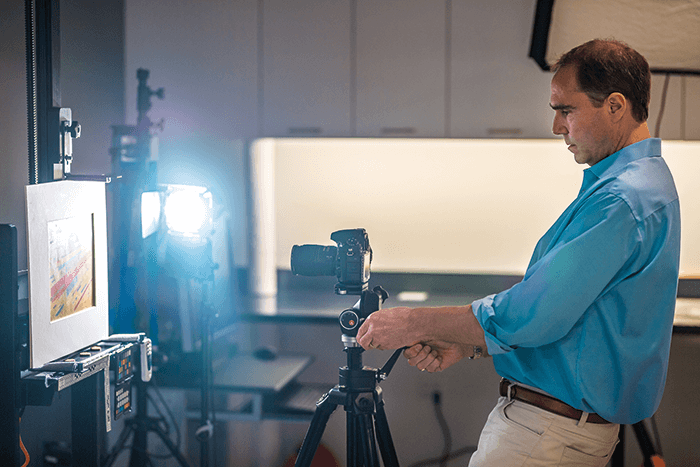
In 2000, I left the Clark and established the first private fee-per-service conservation science laboratory in the US. The firm, Orion Analytical, continued to serve conservators, museums, and the FBI – and soon expanded its clientele to include manufacturers, collectors, law firms, and insurance companies. The materials Orion studied were very diverse – works of art and cultural property (from ancient Egyptian artefacts to contemporary art), architectural finishes, consumer product packaging and sealants… and contaminates on gyroscopes used for guided missile systems! The technical and analytical tools Orion used were diverse, too. Analysis of art can be as “multi-layered“ as the objects we investigate. Conservation scientists generally begin analyses using different wavelengths of energy, from x-rays to infrared, to visualize the composite structure of works and the distribution of materials based on their average atomic mass, visible fluorescence, color, or infrared absorbance. We then use stereomicroscopes to study the surface of objects. Stereomicroscopes are common, but ours are not mounted on the traditional stands you would see in a biological lab – they are mounted on gantries that allow them to be suspended over works of art, and articulated on stands to examine vertical surfaces and three-dimensional objects. These microscopes allow us to look at the general construction and condition of objects, and to look for evidence that works have been intentionally altered to impart a false appearance of age (for example, whether cracks are real, or have been drawn on with a needle or a pencil). Further, stereomicroscopes also allow us to select microscopic areas for in-situ analyses and to remove samples for other analyses. Conservation scientists use non-invasive techniques whenever possible, but samples are usually required to examine layer structure and to identify pigments, polymers, and other organic materials. The good news is that most samples are microscopic in size; for example, the optimal sample size for FTIR microscopy is about 35 µm across, meaning that one could fit several hundred samples on the head of a pin. Further, a single sample often can be used for multiple analyses – SEM-XEDS to map its elemental distribution, a combination of FTIR and Raman to identify its molecular composition – and more. In the same way that a cross-section view of the Grand Canyon tells more about geographic chronology and weathering than does a bird’s eye view, cross-section samples from objects allow us to see and analyze the individual layers used to make works of art, and evidence of the passage of time. For example, fluorescence microscopy of cross-sections helps to elucidate oxidation, weathering between layers and the buildup of grime and contaminants. This systematic use of particle, elemental, and molecular analyses provides information on different physical, optical, and chemical features of materials. Used in combination, these analyses allow us to identify hundreds of thousands of materials, from an ancient pigment or alloy, to natural fibers, to modern synthetic polymers.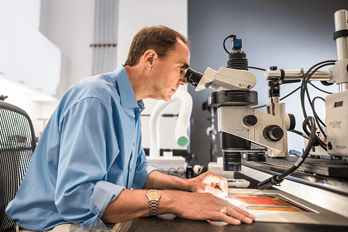
Making a mark
In 2016, Sotheby’s acquired Orion and hired me as its first Director of Scientific Research. Orion had built an unblemished reputation for art analysis, and consulted on the highest-profile art forgery cases between 2000 and 2016. My laboratory at Sotheby’s is the first of its kind in the art industry – in any auction house. To put this into historical perspective, my profession traces its roots to the 1920s, Sotheby’s to 1744! My aim at Sotheby’s is to integrate art conservation science into the day-to-day work of the auction house. In the same way that museum laboratories support the work of curators and conservators, my team supports the work of Sotheby’s researchers and specialists. On any given day, we help our colleagues see hidden parts of works, identify materials and techniques used to create works, and to provide investigative leads that inform the attribution process. The College Art Association codified standards and guidelines for authentication and attribution in 2009, when they identified three essential components – what I liken to a “three-legged stool.” The first essential component is analysis of style by a connoisseur, the only expert who can offer an affirmative attribution or authentication of a work of art – a scientist cannot do that. The second element is the documented history of the work, otherwise known as provenance. The third element is technical and scientific examination to determine whether the physical substance of the work is consistent with its attribution and provenance – what I and my team at Sotheby’s do – like umpires, we call balls and strikes. The laboratory at Sotheby’s includes the techniques I used at Orion, and more. Some of the instruments we use at Sotheby’s also work in the field – anywhere we have electricity or a battery pack. For example, we are the first conservation science laboratory in the world to use a new generation of FTIR microscope that can be hand-carried anywhere – and used without liquid nitrogen. Such instruments make it possible for us to characterize materials on-site, without delay. We have also invested in other cutting-edge instrumentation, including scanning x-ray fluorescence spectrometry (MA-XRF), a technique that scans works of art and produces maps of elemental distribution, often revealing restoration and hidden changes in paintings and other cultural property. Our next purchase likely will be a portable Raman microscope that is sensitive enough to detect materials we see using our laboratory-based microscope, like modern synthetic pigments. My current laboratory is my third, and my favorite! I am chief science officer at Sotheby’s, the largest revolving collection of art in the world – and it is an absolute joy!Newsletters
Receive the latest analytical science news, personalities, education, and career development – weekly to your inbox.


