Conexiant
Login
  • The Analytical Scientist
  • The Cannabis Scientist
  • The Medicine Maker
  • The Ophthalmologist
  • The Pathologist
  • The Traditional Scientist
The Analytical Scientist
  • Explore

    Explore

    • Latest
    • News & Research
    • Trends & Challenges
    • Keynote Interviews
    • Opinion & Personal Narratives
    • Product Profiles
    • App Notes

    Featured Topics

    • Mass Spectrometry
    • Chromatography
    • Spectroscopy

    Issues

    • Latest Issue
    • Archive
  • Topics

    Techniques & Tools

    • Mass Spectrometry
    • Chromatography
    • Spectroscopy
    • Microscopy
    • Sensors
    • Data & AI

    • View All Topics

    Applications & Fields

    • Clinical
    • Environmental
    • Food, Beverage & Agriculture
    • Pharma & Biopharma
    • Omics
    • Forensics
  • People & Profiles

    People & Profiles

    • Power List
    • Voices in the Community
    • Sitting Down With
    • Authors & Contributors
  • Business & Education

    Business & Education

    • Innovation
    • Business & Entrepreneurship
    • Career Pathways
  • Events
    • Live Events
    • Webinars
  • Multimedia
    • Video
Subscribe
Subscribe

False

The Analytical Scientist / Issues / 2020 / Oct / Close to the Edge
Microscopy

Close to the Edge

New single-molecule microscopy technique lets researchers see lipid membranes in unique detail

By Lauren Robertson 10/12/2020 1 min read

Share

Using traditional techniques, such as MS, it’s difficult for scientists to image cell membranes without destroying them in the process. Now, researchers have developed a new system – single-molecule orientation localization microscopy (SMOLM) – that is not only nondestructive, but provides a higher-resolution view of lipid membranes than previously possible (1).

The clue is in the name; SMOLM visualizes the orientation and location of fluorescent probes within the cell membrane. “Any fluorescent probe can be influenced by intermolecular forces from surrounding molecules,” says Matthew Lew, principal investigator. “By measuring a single fluorophore’s orientation within the membrane, we realized we could sense its local environment – an ordered, solid-like lipid membrane will constrain it, whereas a liquid-like one leaves it free to rotate.”

Importantly, the team used lipophilic fluorophores that only transiently attach to lipid bilayers to capture a series of “blinking” images. By collecting hundreds of thousands of frames, they could resolve the lipid membrane’s compositional features.

The researchers then used self-made “lenses” – which they call engineered phase masks (2) – to bend the light within a polarization-sensitive fluorescent microscope. “These masks created images with special multi-spot shapes on our camera,” says Lew. “By measuring the relative brightness of the spots, we can identify the 3D orientation and wobble (or rotational movement) of single fluorescent molecules.” Finally, their image analysis algorithm (3) interprets the data and estimates molecular position, orientation, and wobble across the whole membrane.
 

(Left) Measured 3D orientation (out-of-plane angle) and wobble (rotational motion) of single Nile red lipophilic probes in a lipid membrane with different levels of cholesterol using SMOLM. (Right) SMOLM image shows lipid nanodomains (yellow: ordered phase; blue: disordered phase) within a 3.6×3.6 µm2 region of the membrane. Credit: Jin Lu.

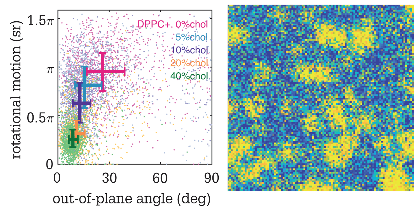
“We’ve demonstrated the feasibility of using SMOLM to resolve lipid nanodomains that are smaller than the diffraction limit of light,” says Jin Lu, lead author of the paper. “Further to this, our method can track enzyme-mediated changes in lipid composition within single domains – a feat that cannot be achieved by conventional methods.”

The team have so far only demonstrated their technique in a model lipid bilayer, but will soon perform further studies in the plasma membranes of living cells.

Newsletters

Receive the latest analytical science news, personalities, education, and career development – weekly to your inbox.

Newsletter Signup Image

References

  1. Jin Lu et al., Angew Chem Int Ed, 59, 17572 (2020). DOI: 10.1002/anie.202006207
  2. O Zhang et al., Appl Phys Lett, 113, 031103 (2018). DOI: 10.1063/1.5031759
  3. H Mazidi et al., IEEE 16th International Symposium on Biomedical Imaging, 325 (2019). DOI: 10.1109/ISBI.2019.8759444

About the Author(s)

Lauren Robertson

By the time I finished my degree in Microbiology I had come to one conclusion – I did not want to work in a lab. Instead, I decided to move to the south of Spain to teach English. After two brilliant years, I realized that I missed science, and what I really enjoyed was communicating scientific ideas – whether that be to four-year-olds or mature professionals. On returning to England I landed a role in science writing and found it combined my passions perfectly. Now at Texere, I get to hone these skills every day by writing about the latest research in an exciting, creative way.

More Articles by Lauren Robertson

False

Advertisement

Recommended

False

False

The Analytical Scientist
Subscribe

About

  • About Us
  • Work at Conexiant Europe
  • Terms and Conditions
  • Privacy Policy
  • Advertise With Us
  • Contact Us

Copyright © 2025 Texere Publishing Limited (trading as Conexiant), with registered number 08113419 whose registered office is at Booths No. 1, Booths Park, Chelford Road, Knutsford, England, WA16 8GS.