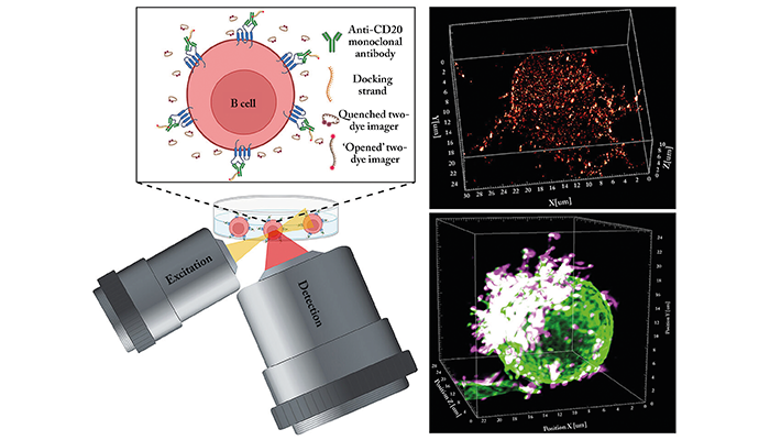
Mode of action of the new microscopy method LLS-TDI-DNA-PAINT. On the top right, the RTX antibody was visualised on a Raij-B cell: it is easy to see how it links the CD20 molecules in the membrane. Bottom right: the hedgehog-shaped appearance of a living Raji B cell after the antibody has bound. The surface protein CD45, which is homogeneously distributed on the cell surface, is also labelled in green.
Credit: Arindam Ghosh / University of Wuerzburg
Researchers have developed a super-resolution microscopy method that reveals how therapeutic antibodies target and destroy cancerous B cells with molecular precision. The technique, called LLS-TDI-DNA-PAINT, provides 3D visualizations of antibody–cell interactions, challenging current classifications of therapeutic antibodies and paving the way for improved immunotherapies.
The study, conducted at Julius-Maximilians-Universität (JMU) Würzburg, focuses on the action of antibodies that bind to the CD20 protein on the surface of B cells, a common target in blood cancers such as chronic lymphocytic leukemia and Burkitt’s lymphoma. These antibodies trigger immune responses that lead to the destruction of cancer cells.
“We can now observe how effectively the antibodies work and thus contribute to the development of improved therapies,” said Professor Markus Sauer, lead researcher and biophysicist at JMU, in a press release.
The researchers used the new method to study the effects of four therapeutic antibodies (RTX, OFA, OBZ, and 2H7) on Raji B cells – a model cell line derived from a Burkitt's lymphoma patient. Their experiments showed that all four antibodies cluster CD20 molecules in specific locations on the cell membrane, particularly on micrometer-long structures called microvilli. This crosslinking polarizes the cell membrane, stabilizing the microvilli on one side of the cell and giving it a hedgehog-like appearance.
“The hedgehog shape makes the B cells appear as if they want to form an immunological synapse with another cell,” said Arindam Ghosh, the study’s first author. This polarization may help activate other immune cells, such as macrophages and natural killer cells, although further research is needed to confirm this.
The findings also challenge the current classification of therapeutic antibodies into Type I and Type II, which are thought to have distinct mechanisms of action. “The previous classification of therapeutic antibodies into types I and II can no longer be maintained,” explained Ghosh, as the study showed similar crosslinking behaviors among all tested antibodies, regardless of type.
The LLS-TDI-DNA-PAINT method allows researchers to observe antibody interactions with tumor cells in both fixed and living states, providing unprecedented insights into their mechanisms of action. The team hopes that these observations will lead to the development of more effective immunotherapies that harness the immune system to fight cancer more efficiently.
Newsletters
Receive the latest analytical science news, personalities, education, and career development – weekly to your inbox.


