Metabolism Activates a Molecular Glue
A multiplexed MS screen identifies a DCAF11 molecular glue that requires glutathionylation before it can drive DDX18 degradation.
A multiplexed mass spectrometry screen has identified a molecular glue that requires metabolic activation, showing that protein degradation can depend on metabolic context while extending discovery to underused E3 ligases.
The platform immobilizes several E3 ligases on beads before exposing them to cell lysate and pooled small molecules. Compounds that increase a ligase’s affinity for a cellular protein draw that protein into the complex, where LC–MS can identify it. Screening seven E3 ligases against 5,000 compounds pointed to recruitment of the RNA helicase DDX18 by DCAF11 and eventually traced the interaction to M12.
In cells, M12 drove DCAF11-dependent degradation of DDX18, but the interaction disappeared when the proteins were tested in purified form. That discrepancy shifted attention to the compound itself. Mass spectrometry showed that M12 had been glutathionylated in the lysate, generating glutathione-modified M12 (GSH-M12) and revealing that metabolic modification was required for degradation.
“This was a huge surprise, and it is the first observation of a molecular glue that has been activated metabolically by glutathionylation,” said co-first author Franziska Wachter in a Dana-Farber press release. Cryo-electron microscopy showed the glutathione group occupying a conserved binding pocket in DCAF11, while the exposed M12 portion remodeled the ligase surface to recruit DDX18.
The chemistry also proved adaptable. Using GSH-M12 as a DCAF11-recruiting element, the researchers built bifunctional degraders targeting proteins including SMARCA2, WEE1, and CDK7. This suggests that metabolic activation could become a design feature rather than simply an unexpected property of one glue.
The authors now plan to examine how glutathione and glutathione S-transferase levels across cancer cells influence M12 activity, while extending the screening strategy to additional ligases and chemical libraries.
“This novel platform is an exciting scalable approach to the discovery of molecular glues,” said co-senior author Eric Fischer.
Freezing the Metabolome in Place
In-source freezing improves IR-MALDESI spatial resolution while suppressing post-harvest metabolic changes in fresh leaf tissue.
Freezing freshly harvested leaf tissue directly inside an IR-MALDESI source has improved spatial resolution while suppressing metabolic changes that can occur between harvesting and mass spectrometry imaging (MSI).
Infrared matrix-assisted laser desorption electrospray ionization (IR-MALDESI) can image fresh leaves with little preparation by penetrating the cuticle and using endogenous water as a matrix. The challenge is that harvesting can trigger rapid stress responses that alter the metabolite distributions MSI is intended to capture.
To limit those changes, the researchers mounted freshly harvested white clover leaves on carboxymethylcellulose gel and froze them to −10 °C on the instrument’s cooled stage. Keeping the tissue frozen throughout imaging also avoided thawing, which can rupture water-rich plant cells and redistribute metabolites.
Freezing also changed how the laser interacted with the tissue. Average ablation-spot diameter fell from 65.4 to 43.9 micrometers, providing an approximately 1.5-fold improvement in spatial resolution with a conventional 2.97-micrometer infrared laser. The smaller spots produced slightly lower overall ion abundance, reflecting the trade-off between spatial resolution and the amount of material sampled.
The metabolic profiles suggest that freezing also preserved a closer snapshot of the leaf at harvest. In fresh tissue, ratios of cinnamic acid and p-coumaric acid to phenylalanine increased between sequentially analyzed leaflets, consistent with continued activity in the stress-responsive phenylpropanoid pathway. That pattern was absent from frozen samples, supporting metabolic quenching during analysis.
The authors suggest that in-source freezing could improve the biological accuracy of IR-MALDESI plant imaging while providing finer spatial detail. Testing thicker and more structurally diverse tissues should help establish how broadly the approach can be applied and how the improved resolution affects sensitivity across metabolite classes.
Which PASEF Works Best for Metaproteomics?
A five-way benchmark identifies DIA- and Slice-PASEF as the strongest overall performers for gut metaproteomics.
A five-way comparison of mass spectrometry acquisition strategies has identified DIA- and Slice-PASEF as the strongest overall performers for gut metaproteomics, improving low-abundance protein detection and functional coverage in complex fecal samples.
The study compared five parallel accumulation–serial fragmentation (PASEF) modes: data-dependent acquisition (DDA), data-independent acquisition (DIA), Slice-, Synchro-, and midia-PASEF. Defined amounts of two bacterial proteomes provided a controlled benchmark for sensitivity, quantitative accuracy, and species-abundance scaling against the more complex fecal background.
DIA-based approaches generally recovered more microbial and host proteins than conventional DDA-PASEF and retained better coverage at low abundance. DIA- and Slice-PASEF also supported taxonomic and functional assignments with more peptide and protein evidence.
“Metaproteomics allows us to see what microbial communities are actually doing, rather than simply which microbes are present,” said first author Feng Xian in the university’s press release. “By identifying the most suitable analytical strategies, we hope to make future microbiome research more sensitive, reproducible, and accessible across medicine, environmental science, and biotechnology.”
The two leading approaches emphasized different trade-offs. Slice-PASEF produced the tightest abundance-ratio distributions and most consistent functional quantification, while DIA-PASEF offered a more balanced combination of depth, precision, and throughput. Extending LC gradients from five to 22 minutes substantially improved coverage, with smaller gains between 22 and 45 minutes.
Applied to a mouse model of intestinal injury, both methods captured closely aligned microbial and host responses during inflammation and recovery, showing that the analytical gains translated into consistent biological interpretation.
The authors suggest that the benchmark can help researchers match acquisition strategy to study design as metaproteomics moves toward larger clinical cohorts.
“Developing better technologies is essential if we want to answer the next generation of questions in microbiome research,” said David Gomez-Varela.
How Gut Microbes Limit Fat Absorption
GC-MS and lipidomics reveal how microbial colonization alters bile composition to retain more dietary lipid in the intestine.
Stable-isotope tracing has shown how the gut microbiota can restrict dietary lipid absorption in mice, linking microbial colonization to changes in bile composition that retain more ingested lipid in the intestine.
The study used deuterium-labelled fatty acid and triglyceride tracers to follow dietary lipid uptake in germ-free mice and animals carrying either a defined or conventional gut microbiota. Gas chromatography–mass spectrometry tracked the labels through intestinal contents, plasma, bile, and peripheral tissues. Colonized mice retained more labelled fatty acids in the gut while less reached the circulation and adipose tissue, pointing to reduced absorption rather than impaired triglyceride breakdown.
Lipidomics narrowed the effect to phosphatidylcholine, a bile component needed for micelle formation. Levels fell with microbial colonization, while supplying additional phosphatidylcholine roughly doubled labelled fatty-acid levels in plasma and liver after one hour.
Further isotope experiments linked that loss to phospholipase A1 activity in bile. High-resolution MS showed faster conversion of labelled phosphatidylcholine to lysophosphatidylcholine in colonized mice, while proteomic and bile-acid analyses connected the effect to host regulation rather than direct microbial degradation.
The resulting pathway links microbial sensing through MYD88 to reduced hepatic CYP7B1 activity and increased taurocholate. Taurocholate then promotes phosphatidylcholine degradation, limiting micelle formation and ultimately reducing intestinal lipid uptake.
By tracing the effect to bile phosphatidylcholine turnover, the study identifies a specific host pathway through which the microbiota can influence lipid uptake. The next question is whether the same mechanism operates strongly enough in humans to offer a practical metabolic target.
(Mass) Spectacular and Strange
A Gene Gun for Greener Meat
A gene gun, gold particles, and lettuce chloroplasts have opened a new route to plant-grown muscle protein.
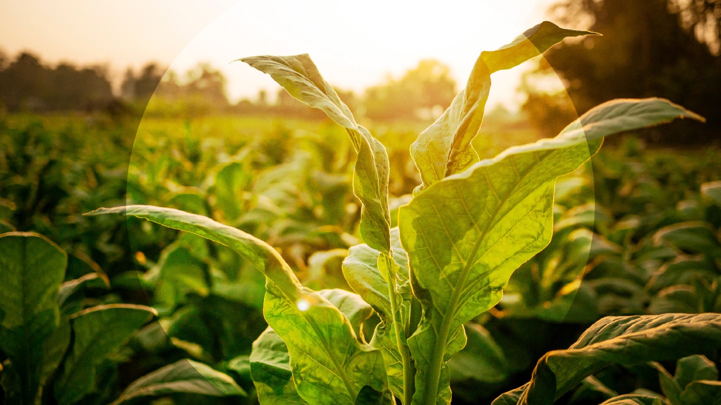
A new study led by researchers at Imperial College London and Kyomei has shown that tobacco and lettuce chloroplasts can be engineered to produce porcine myoglobin. The heme-binding muscle protein helps give meat its red color, heme-associated iron, and metallic, umami flavor. The team used biolistic transformation to fire DNA-coated gold particles into plant tissue, inserting myoglobin genes into the chloroplast genome.
Chloroplasts are attractive targets because they retain bacterial-like gene-expression machinery and occur in many copies per plant cell. The transformed tobacco and lettuce plants grew to maturity, flowered, and produced seeds that inherited the engineered trait.
The team confirmed myoglobin accumulation using SDS-PAGE and immunoblotting, then purified the protein from tobacco leaves for further analysis. LC-MS showed a protein mass consistent with myoglobin after removal of its initiator methionine, a common chloroplast modification, and also detected heme b in the sample. Heme binding remained incomplete, however, suggesting that heme availability may still limit production.
Yields reached around 2.8 percent of total soluble protein in tobacco and 1.5 percent in lettuce, equivalent to approximately 800 mg of myoglobin per kilogram dry weight in both plants. The work remains a proof-of-concept, with heme availability still a likely bottleneck, but it points to a possible route for growing meat-relevant proteins in crops.
“Here we show that plants can be engineered to produce the animal protein myoglobin in their chloroplasts,” said Alexia Groff in the Frontiers press release. “This could provide a more sustainable way to produce an important ingredient for plant-based meat products.”
Newsletters
Receive the latest analytical science news, personalities, education, and career development – weekly to your inbox.