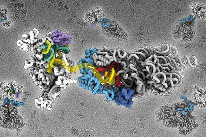
Researchers have developed a nuclear magnetic resonance (NMR) spectroscopy method that can examine proteins inside intact mitochondria. The approach provides detailed information about protein structure, interactions, and stability within these cellular compartments.
Mitochondrial dysfunction has been linked to cancer, inflammation, aging, and neurodegenerative diseases. However, studying proteins in purified laboratory solutions may not accurately reflect how they behave inside mitochondria.
The researchers isolated mitochondria from rat liver and pig brain tissue. They produced selected proteins in Escherichia coli, labeled them with NMR-detectable isotopes, and delivered them into the mitochondria using electroporation.
Tests of membrane potential and enzyme activity indicated that the mitochondria remained intact and functional after the procedure. Control experiments also confirmed that most of the measured signals came from proteins inside the mitochondria rather than proteins that had leaked into the surrounding solution.
The team used nitrogen-15 and fluorine-19 labeling to study alpha-synuclein, a protein associated with Parkinson’s disease, along with three folded proteins: GB1, ubiquitin, and KH1. Selective labeling helped reduce overlapping signals, while fluorine-19 produced sensitive measurements with little interference from naturally occurring biological molecules.
The results indicated that alpha-synuclein remained largely disordered inside mitochondria. Changes in its NMR signals showed that the protein’s N-terminal region interacted with mitochondrial membranes. The A30P variant, which is associated with inherited Parkinson’s disease, showed weaker membrane interactions than the unmodified protein.
The researchers also examined the effects of methionine oxidation, a chemical modification associated with oxidative stress. Oxidation reduced the interaction between alpha-synuclein’s N-terminal region and mitochondrial membranes. This finding may help researchers investigate how oxidative stress changes protein behavior within mitochondria, although the study did not assess disease progression or patient outcomes.
Further experiments examined the stability of GB1. The protein was more stable inside mitochondria than in a laboratory buffer, possibly because of molecular crowding and interactions with other mitochondrial components.
The researchers also measured the binding between two copies of a GB1 variant. Its binding affinity was approximately twice as high inside mitochondria as it was in buffer, showing that the organelle’s internal environment can influence protein-protein interactions.
The method could help researchers study proteins within a more biologically relevant setting and investigate molecular processes associated with mitochondrial disease. However, the experiments used isolated mitochondria at temperatures below normal physiological conditions. Samples also remained viable for a limited period, restricting the study of slower processes.
Further work is needed to improve measurement sensitivity, extend sample viability, and assess additional proteins and disease models.
Newsletters
Receive the latest analytical science news, personalities, education, and career development – weekly to your inbox.