“My research focuses on the development and adaptation of remote sensing imaging techniques to study works of art in support of conservation and art history,” says John Delaney, Senior Imaging Scientist at the National Gallery of Art in Washington, DC. He is also a Research Professor in the Department of Electrical and Computer Engineering at George Washington University. The job is, he says,” a dream come true.” John, who joined the National Gallery of Art in 2007, was previously the Chief Scientist for the Advance Sensors Business Unit of Airborne ISR Systems at Goodrich Corporation.
Can the pigments and paint binders used in illuminated manuscripts be mapped in situ?
An illumination from a choir book commissioned by the Camaldolese monks of Santa Maria degli Angeli in Florence at the end of the fourteenth century. This commission includes some of the most beautifully illuminated volumes of the early Italian Renaissance. Contributing significantly to the production of these books was Lorenzo Monaco, known as one of the greatest panel painters of the time, and an accomplished illuminator of manuscripts. The illumination studied and discussed here (see figure) represents a bearded prophet with his hands raised in prayer, within a blue and green initial E decorated with filigree ornaments, blue and yellow circular ‘gems’, red-glazed gilt diamonds, and colorful foliage. The background is gold leaf.
- Manuscript illuminations are extremely sensitive to physical handling, to environmental changes and to light.
- The collection of even a small sample would mar the miniature’s appearance, ruling out analytical chemical methods such as HPLC and GC-MS.
- While site-specific in situ analytical tools, including X-ray fluorescence and Raman spectroscopy, do not require a sample, there is growing interest in obtaining chemical information over an entire art object.
Visible and near infrared (NIR) reflectance imaging spectroscopy is the collection of contiguous calibrated spectral images to provide the reflectance spectrum for each pixel of the scene. The method utilizes information from both electronic transitions in the visible and near infrared, as well as vibrational features from various organic and inorganic chemical groups, namely hydroxyl, carbonate, methylenic and amide groups. It was developed to remotely map and identify minerals and vegetation but applications have ranged from the study of planets to process modeling in the pharmaceutical industry.
The sensitivity of manuscript illuminations requires the optimization of hyperspectral camera systems operating from 400 to 2500 nm with a few nm sampling to conduct such studies. This is achieved by using high-sensitivity and low-noise visible and infrared focal planes that were originally developed for scientific and military applications. Once the image cubes (see figure) whose third dimension is spectral, are collected and calibrated, the hundreds of images are processed via an algorithm developed for clustering and setting apart the basis set of reflectance spectra which describe the art work. This method relies on both principal component analysis and convex geometry. The resulting spectral ‘endmember’ can be mapped using one of several algorithms including the ‘spectral angle mapper’, which identifies all the pixels in the cube whose spectra match that of the reference endmenber to within a fixed tolerance value. Identification of the artist’s material is then done in two ways: comparison with reflectance spectral databases, and by noting characteristic vibrational features such as position and slope of electronic transitions.
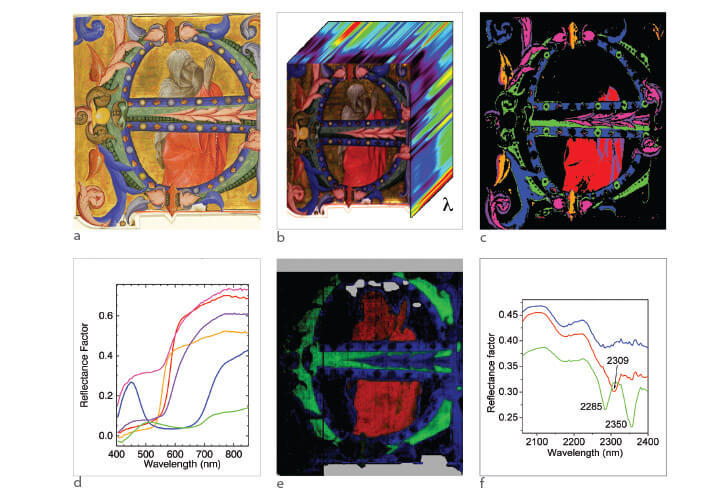
The analysis of the hyperspectral Vis-NIR cube and other analytical data revealed that the mineral azurite was used for the green portion of the initial, while blue areas were painted using two grades of ultramarine blue. The orange leaves were painted with red lead and the pink leaves with an insect-derived red dye. The spectral data also showed that the red robe of the Prophet was painted using vermilion and glazed with the red dye. Analysis of the NIR cube also allowed a new insight into Monaco’s painting technique, specifically the use of a fat-containing paint binder (likely egg yolk) only for certain compositional elements of this manuscript leaf – the figure of the prophet, but not the decorated initial. The use of any fat-containing binder for manuscript illumination is surprising in itself since egg white and gum Arabic (based on proteins and polysaccharides) are historically considered to be the binders preferred by illuminators. This shows how the use of reflectance imaging spectroscopy in the visible and NIR opens the opportunity for conservation scientists, conservators and art historians to explore further the painting techniques of Lorenzo Monaco and of other illuminators.
Please read the other articles in this series:
The Science of Art
Deconvoluting the Creative Process
The Stories That Colours Tell
Understanding Ancient Prescriptions
Newsletters
Receive the latest analytical science news, personalities, education, and career development – weekly to your inbox.

References
- Ricciardi et al. Angew Chem Int Ed Engl. 2012 Jun 4;51(23):5607-10


