Using traditional techniques, such as MS, it’s difficult for scientists to image cell membranes without destroying them in the process. Now, researchers have developed a new system – single-molecule orientation localization microscopy (SMOLM) – that is not only nondestructive, but provides a higher-resolution view of lipid membranes than previously possible (1).
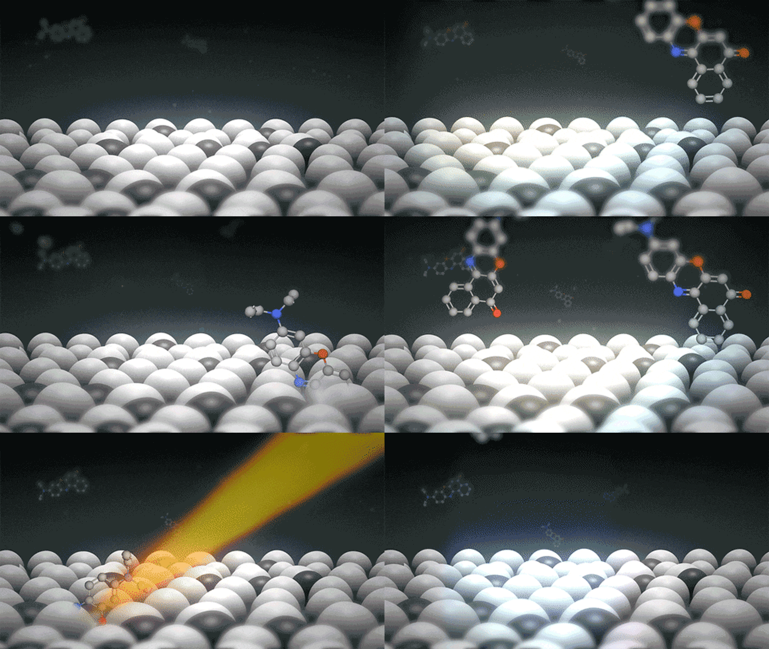
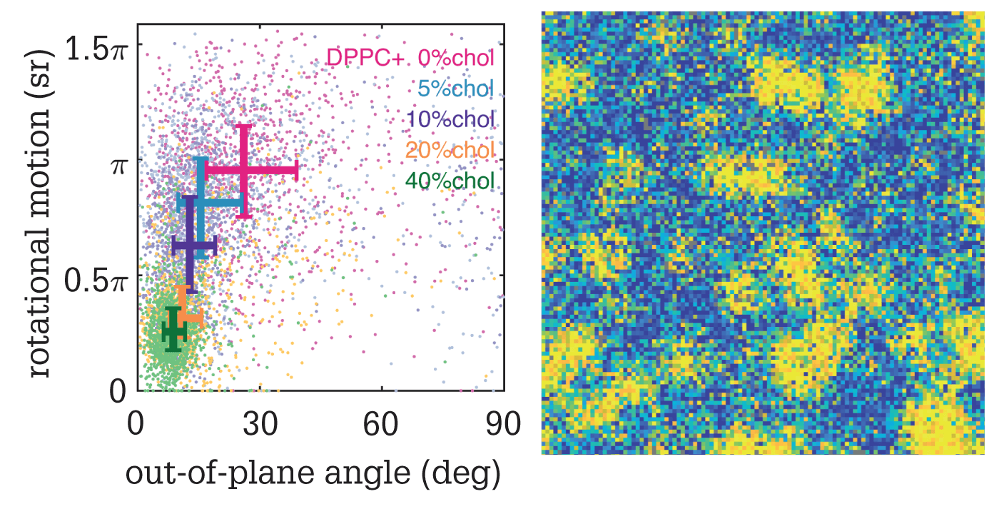
The clue is in the name; SMOLM visualizes the orientation and location of fluorescent probes within the cell membrane. “Any fluorescent probe can be influenced by intermolecular forces from surrounding molecules,” says Matthew Lew, principal investigator. “By measuring a single fluorophore’s orientation within the membrane, we realized we could sense its local environment – an ordered, solid-like lipid membrane will constrain it, whereas a liquid-like one leaves it free to rotate.”
Importantly, the team used lipophilic fluorophores that only transiently attach to lipid bilayers to capture a series of “blinking” images. By collecting hundreds of thousands of frames, they could resolve the lipid membrane’s compositional features.
The researchers then used self-made “lenses” – which they call engineered phase masks (2) – to bend the light within a polarization-sensitive fluorescent microscope. “These masks created images with special multi-spot shapes on our camera,” says Lew. “By measuring the relative brightness of the spots, we can identify the 3D orientation and wobble (or rotational movement) of single fluorescent molecules.” Finally, their image analysis algorithm (3) interprets the data and estimates molecular position, orientation, and wobble across the whole membrane.

“We’ve demonstrated the feasibility of using SMOLM to resolve lipid nanodomains that are smaller than the diffraction limit of light,” says Jin Lu, lead author of the paper. “Further to this, our method can track enzyme-mediated changes in lipid composition within single domains – a feat that cannot be achieved by conventional methods.”
The team have so far only demonstrated their technique in a model lipid bilayer, but will soon perform further studies in the plasma membranes of living cells.
References
- Jin Lu et al., Angew Chem Int Ed, 59, 17572 (2020). DOI: 10.1002/anie.202006207
- O Zhang et al., Appl Phys Lett, 113, 031103 (2018). DOI: 10.1063/1.5031759
- H Mazidi et al., IEEE 16th International Symposium on Biomedical Imaging, 325 (2019). DOI: 10.1109/ISBI.2019.8759444


