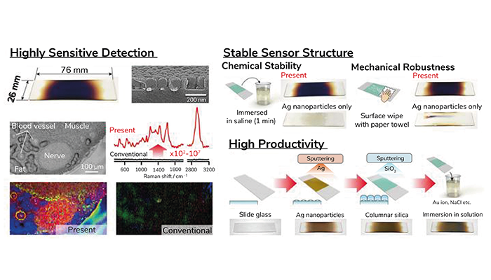
Highly sensitive molecular detection with chemical stability and mechanical robustness through long-range molecule-plasmon interactions.
Credit: Takeo Minamikawa, Reiko Sakaguchi, Yoshinori Harada, Hiroki Tanioka, Sota Inoue, Hideharu Hase, Yasuo Mori, Tetsuro Takamatsu, Yu Yamasaki, Yukihiro Morimoto, Masahiro Kawasaki, and Mitsuo Kawasaki
Researchers have developed a nanostructured surface that enables long-range signal enhancement for fluorescence and Raman spectroscopy, potentially enabling highly sensitive, non-invasive biosensing and molecular analyses.
The new technique, developed by a team of researchers at Osaka University led by Takeo Minamikawa, combines a column-structured silica (CSS) layer with silver nanoislands (AgNIs). The CSS layer, over 100 nm thick, physically separates the AgNIs from analytes, preventing mutual degradation while maintaining significant signal amplification.
In testing, the device – dubbed a remote plasmonic-like enhancement (RPE) plate – achieved Raman signal enhancement up to 107 times and fluorescence increases up to 102 times compared with traditional methods.
The fabrication process begins with AgNIs deposited on glass through sputtering, followed by a CSS layer to shield the AgNIs. A gold(I)/halide treatment optimizes the plasmonic response, and testing – using energy-dispersive X-ray spectroscopy (EDS) and scanning electron microscopy (SEM) – confirmed that analytes remain isolated on the silica surface.
"We demonstrated that the range of influence of plasmons in metals can exceed 100 nanometers, far beyond what conventional theory predicted," Minamikawa explained, in a press release.
The team validated the technology through two key applications. In biosensing, enhanced fluorescence signals revealed detailed Ca²⁺ signaling in live HeLa cells with minimal disruption. For tissue imaging, RPE-enhanced Raman imaging produced distinct spectral signatures from biological tissues, enabling high-resolution mapping.
Senior author Mitsuo Kawasaki noted: "The chemical stability and mechanical robustness of our substrates make them suitable for a wide range of applications, including environmental pollutant detection or medical diagnosis."


