Subscribe to Newsletter
Techniques & Tools Microscopy

Image of the Month: Doubling Up

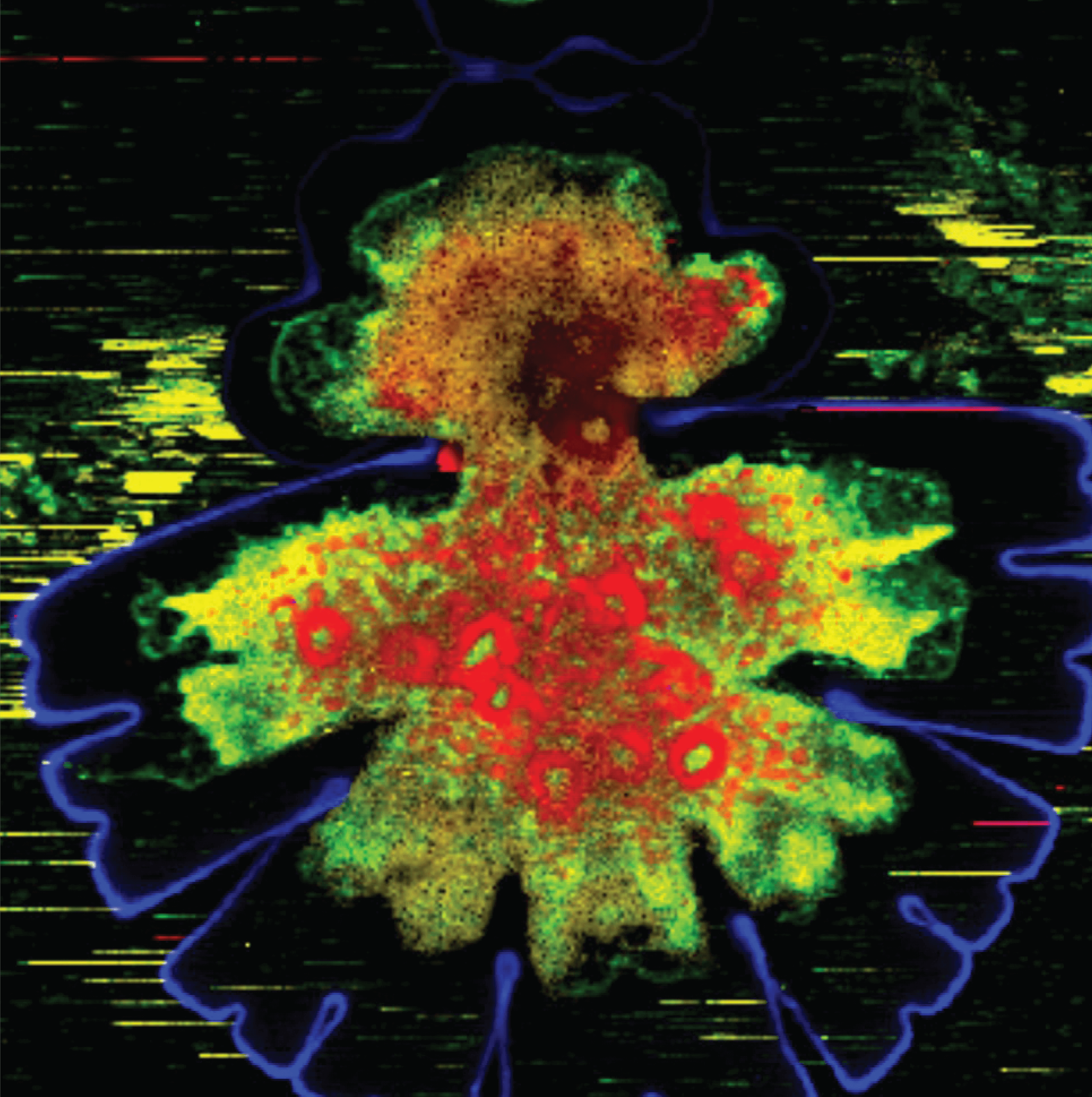
Micrasterias denticulata is a green alga found in acidic-to-neutral fresh waters and sphagnum bogs. This confocal Raman micrograph shows the unicellular organism undergoing cell division; the cell wall is shown in blue, starch in red, and proteins, pectins and fats in green and yellow. The entire image width is a mere 160 μm.

Credit: Dr Notburga Gierlinger, BOKU; licensing information available here .

Receive content, products, events as well as relevant industry updates from The Analytical Scientist and its sponsors.
Stay up to date with our other newsletters and sponsors information, tailored specifically to the fields you are interested in

When you click “Subscribe” we will email you a link, which you must click to verify the email address above and activate your subscription. If you do not receive this email, please contact us at [email protected].
If you wish to unsubscribe, you can update your preferences at any point.

About the Author
Matt Hallam

I've always wanted a job that fosters creativity - even when I worked on the assembly line in a fish factory. Outside work, I satisfy this need by writing questionable fiction. The venture into science writing was an unexpected departure from this fiction, but I'm truly grateful for the opportunity to combine my creative side with my scientific mind as Editor of The Analytical Scientist.

Register to The Analytical Scientist

Register to access our FREE online portfolio, request the magazine in print and manage your preferences.

You will benefit from:
  • Unlimited access to ALL articles
  • News, interviews & opinions from leading industry experts
  • Receive print (and PDF) copies of The Analytical Scientist magazine

Register