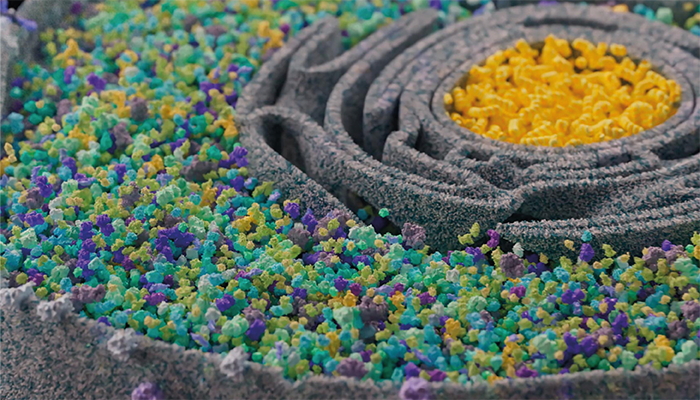
A hustle and bustle like at the Zurich Street Parade: inside a cell, countless different proteins interact with each other around the cell nucleus.
Credit: Cathy Marulli / ETH Zurich
A team of researchers at ETH Zurich have developed a high-throughput workflow known as serial ultrafiltration combined with limited proteolysis coupled to mass spectrometry (FLiP-MS) to systematically monitor protein complex dynamics and thereby generate a library of peptide markers specific to changes in protein-protein interactions (PPIs).
The method works by exposing lysates to a non-specific protease (such as proteinase K), which can only cleave proteins at freely accessible sites and therefore exposes differences between protein structures. This is an evolution of LiP mass spectrometry – a technique developed by Paola Picotti’s team in 2017 that enables the measurement of structural changes between proteins in any biological sample without the need for prior purification.
The team applied FLiP-MS to the Saccharomyces cerevisiae proteome to probe PPIs, identifying markers for over 1,000 proteins, which they used to identify links between the assembly states of complexes such as Spt-Ada-Gcn5 acetyltransferase (SAGA).
They believe that the technology could lead to the development of a library of protein-binding interfaces (PBIs) to better understand the effects of mutations in cancers, and diseases such as Alzheimer’s.
We spoke to authors Paola Picotti, Cathy Marulli and Natalie de Souza to get their insights on the technology, and what they believe their findings could mean for the wider scientific community.
Please can you give us an introduction to LiP mass spectrometry?
Limited proteolysis coupled to mass spectrometry (LiP-MS) is a technology that probes structural changes in proteins between any two conditions of interest. Notably, LiP-MS is applicable in a complex environment such as a cell or tissue lysate and can therefore determine, for thousands of proteins simultaneously, which of them change structure between two conditions. Since protein structure is intimately linked to protein function, identifying proteome-wide changes in structure is a very powerful way to understand functional changes between two conditions. For example, between health and disease or when cells are subjected to DNA replication stress, as we look at in this study.
In brief, LiP-MS works by exposing native lysates for a short time to a non-specific protease (proteinase K), which cleaves flexible and surface accessible residues. Changes in protein structure are reflected in different patterns of proteolysis, since different residues are accessible in different protein conformations. Afterwards, the proteins are denatured, digested to peptides with trypsin, and processed in the standard manner for mass spectrometry. Differential peptide abundances across conditions then pinpoint protein regions changing structure between those conditions, for all detected proteins. LiP-MS generates very rich reports of information on all kinds of structural alterations, ranging from protein-protein binding, protein-small molecule binding, and post-translational modifications, to conformational changes underlying allosteric regulation and enzyme activity changes.
How did you further develop your LiP mass spec approach for this study?
One challenging aspect of the very rich information contained in a LiP-MS dataset is that it isn’t easy to interpret specific LiP changes in molecular terms. In other words, since LiP-MS captures many different types of structural changes, one does not a priori know which ones reflect, for instance, altered protein-protein interactions (PPIs). Changes in PPIs – that is, assembly or disassembly of protein complexes – are particularly interesting, not only because many biological functions are carried out by protein complexes, but also because they are potentially interesting as drug targets. So in this study, we developed a way to disentangle the information in a LiP-MS dataset, in order to determine which LiP peptide changes are reporting on changes in PPIs between two conditions.
Our strategy was to generate an experimental library of peptides that we know are associated with protein-protein binding events, and that could therefore serve as markers of these events. The rationale behind this is rather simple: if we manage to specifically probe structural differences between complex-bound and monomeric forms of proteins, the peptides that indicate those differences can be subsequently used as markers to identify peptides from LiP-MS experiments that reflect changes in protein-protein interactions (PPIs).
Another major challenge was to find a method that efficiently fractionates lysates to separate protein complexes from their monomeric counterparts globally, whilst also producing comparable and high enough protein concentrations in the different fractions, as this is crucial for the limited proteolysis step. Our initial efforts to do this with size-exclusion chromatography (SEC) were somewhat successful, but we concluded that the large dilution factor during SEC was suboptimal for the study of protein complexes. We therefore chose serial ultrafiltration as our fractionation method, which concentrates lysates rather than diluting them. Even though this seemed quite straightforward in principle, it turned out that we need a lot of input material (1 liter of yeast culture per replicate) to reach high enough protein concentrations in all filter fractions. Nevertheless, serial ultrafiltration finally allowed us to efficiently separate protein complexes from their monomeric counterparts and generate high quality LiP readouts reporting on the structural differences between different assembly states.
Did anything surprise you about your findings?
This wasn’t so much a surprise as more of a realization of hope, but one of the aspects of FLiP-MS worth emphasizing is that it makes it possible to look at not only a static protein interactome but a dynamic one, under any desired genetic or environmental perturbation or even both, as we’ve shown here. Since we also have maps of where in any given protein the changes occur, the approach gives us both a birds-eye view of what’s happening to protein complexes in the cell as well as the means to zoom into what’s going on in a specific protein.
Another aspect of the study that was gratifying (though again not really surprising) was to see just how many protein complexes changed upon perturbation. This is not unexpected, but it’s exciting to have an approach that actually allows one to analyze it.
Thinking of the big picture, what is the full potential of your technology?
There is more and more literature showing that PPI networks largely rewire in human diseases, suggesting that targeting those interactions could be a therapeutic strategy. Our current study is in yeast, but can easily be implemented in a human system. Once we’ve done that, FLiP-MS has the throughput and scalability to, in principle, track changing PPIs across the human proteome in patient samples to pinpoint interactions dysregulated in a disease, possibly even in a personalized manner. Furthermore, although the FLiP marker library does not exclusively contain peptides at protein binding interfaces, the markers could be combined with protein structure prediction tools to validate binding interfaces and guide drug design. Finally, what we ourselves are most interested in is that FLiP markers could be used to screen compounds that stabilize or destabilize PPIs; an approach that shows great potential to target proteins currently considered undruggable.


