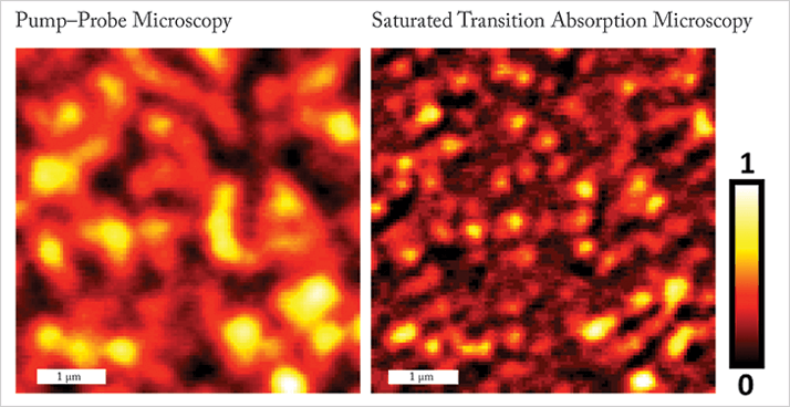
The diffraction limit for non-fluorescent species – the fundamental limit of optical imaging resolution – has been breached. Researchers at Purdue University, Lafayette, Indiana, imaged graphite nanoplatelets about 100 nm wide (see image) (1). Conventional optical microscopes cannot resolve objects smaller than around 300 nm.
The group has developed a new iteration of super-resolution optical microscopy called saturated transient absorption microscopy (STAM). According to Ji-Xin Cheng, an associate professor of biomedical engineering and chemistry at Purdue, the project was initiated a few years ago. “We developed pump-probe microscopy to study nanomaterials, and published our first paper in PRL (2),” he explains. “At that time, we were able to distinguish semiconducting versus metallic nanotubes; however, our resolution was not sufficient to separate adjacent nanotubes. This constraint triggered us to push the limit of resolution in label-free imaging”.
In very simple terms, the addition of a third doughnut-shaped laser to the already complex pump–probe microscopy setup creates a reduced focal centre, which enabled the breakthrough.
The diffraction limit has been broken before, ushering in the advent of super-resolution optical microscopy, but native materials had not been imaged. “Up until now, far-field super-resolution studies have been based on fluorescence in a labeled specimen,” Cheng says. “Ours is based on inherent absorption and is label-free.” In fact, the doughnut shaped laser in the new system is based on that used in stimulated emission depletion (STED) microscopy, developed by Stefan Hell, now director of Max Planck Institute for Biophysical Chemistry in Goettingen, Germany. STED demonstrated the potential of super-resolution microscopy using fluorescent labels back in 1999. But because STAM is label free, the signal is obtained directly from the material itself, providing a more detailed understanding of its nanostructure. It means that STAM can be applied in new areas, including, Cheng says, “the electronic properties in nanodomains of 2D nanomaterials and the properties of single nanostructures in situ, for example, in a solar cell.”
Graphene is an attractive candidate for study using STAM due to its photo-stability, which is a fundamental requirement of the technique. Proteins and lipids are likely targets in the biomedical space.
Cheng and colleagues hope to drive resolution down to 10 nm. “We are not there yet,” he said in a previous interview, “But a few schemes can be applied to further increase the resolution of our system.”
Click here for a video presentation on STED
Newsletters
Receive the latest analytical science news, personalities, education, and career development – weekly to your inbox.

References
- UC4gV2FuZyBldCBhbC4sIOKAnEZhci1maWVsZCBpbWFnaW5nIG9mIG5vbi1mbHVvcmVzY2VudCBz cGVjaWVzIHdpdGggc3ViZGlmZnJhY3Rpb24gcmVzb2x1dGlvbizigJ0gTmF0dXJlIFBob3Rvbmlj cyBkb2k6MTAuMTAzOC9ucGhvdG9uLjIwMTMuOTcgKDIwMTMpLg0KWS4gSnVuZyBldCBhbC4sIOKA nEZhc3QgRGV0ZWN0aW9uIG9mIHRoZSBNZXRhbGxpYyBTdGF0ZSBvZiBJbmRpdmlkdWFsIFNpbmds ZS1XYWxsZWQgQ2FyYm9uIAhOYW5vdHViZXMgVXNpbmcgYSBUcmFuc2llbnQtQWJzb3JwdGlvbiBP cHRpY2FsIE1pY3Jvc2NvcGUs4oCdIFBSTCAxMDUsIDIxNzQwMSAoMjAxMCku


