Credit: Molly M Bechtel, University of California, Davis
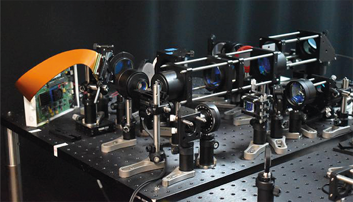
A new two-photon fluorescence microscope developed by researchers at the University of California, Davis, allows for high-speed imaging at cellular resolution, while minimizing potential damage to brain tissue. The new system could provide critical insights into brain function and aid in the study of neurological diseases.
Traditional two-photon microscopy, while effective at imaging deep within brain tissue, is limited by slow imaging speeds and potential photodamage caused by the laser scanning process. The UC Davis team, led by Weijian Yang, addressed these limitations by replacing the conventional point-scanning approach with an adaptive line-excitation technique. This new method illuminates only the regions of interest – specific neurons – rather than scanning the entire field of view, thereby reducing the total laser exposure and associated tissue damage.
“Our new microscope is ideally suited for studying the dynamics of neural networks in real time, which is crucial for understanding fundamental brain functions such as learning, memory and decision-making,” said Yang in a press release. “For example, researchers could use it to observe neural activity during learning to better understand communication and interaction among different neurons during this process.”
Key to this innovation is the use of a digital micromirror device (DMD), which dynamically shapes and directs the laser beam based on the identified regions of interest. This setup allows the system to capture large areas of active neurons in a single pass, significantly increasing the imaging speed – by up to 10 times faster than traditional two-photon microscopy.
The adaptive line-excitation method was tested in vivo on mouse cortex tissue, where it successfully captured calcium signals, which are indicators of neural activity, at a rate of 198 Hz. This is a significant improvement over conventional systems, which are often too slow to capture rapid neuronal events. The study demonstrated that the new method could isolate the activity of individual neurons with high accuracy, even in densely packed neural environments.
The combination of high-speed imaging and reduced laser power not only protects the brain tissue but also enables the system to focus on the most relevant neuronal activity. This selective illumination results in less background noise and clearer images, making it easier to interpret complex neural interactions.
The research team is now working to integrate voltage imaging capabilities into the system, which would allow for even more precise monitoring of neural activity. They also plan to refine the microscope’s design to make it more user-friendly and compact, which would broaden its application in various neuroscience research settings.
The UC Davis team’s work holds promise for a wide range of applications, from studying basic neural processes, such as learning and memory, to investigating the early stages of neurological diseases like Alzheimer’s and Parkinson’s.
Newsletters
Receive the latest analytical science news, personalities, education, and career development – weekly to your inbox.


