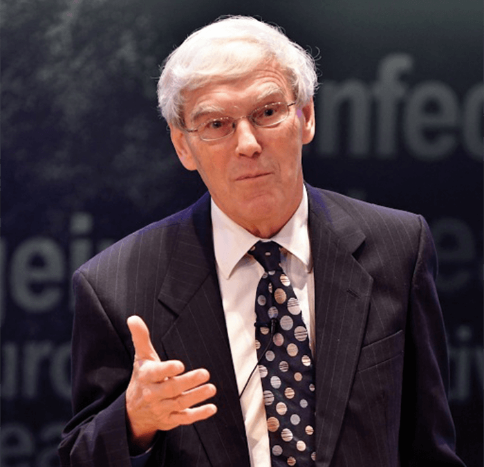
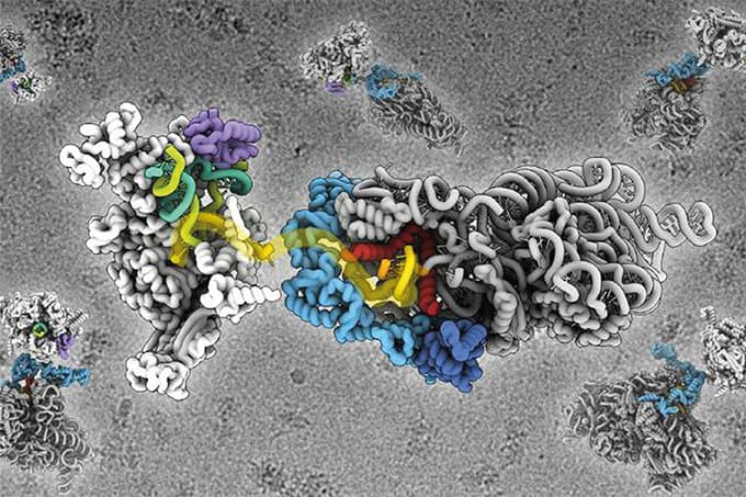
While the world hurries to congratulate the 2018 Nobel Prize winners, we’re still celebrating Richard Henderson, who, along with Jacques Dubochet and Joachim Frank, won the 2017 Nobel Prize in Chemistry “for developing cryo-electron microscopy for the high-resolution structure determination of biomolecules in solution (1).” Why? On September 12, 2018, Henderson officially opened Diamond Light Source’s electron bio-imaging centre (eBIC) in Cambridge, UK. The event coincided with the announcement of a partnership between Diamond and Thermo Fisher Scientific, which adds two new microscopes and professional cryo-EM services specifically for the pharmaceutical industry. The additional capacity makes eBIC one of the largest cryo-EM sites in the world – a true nod to the technology’s fast-growing significance in structural biology. Rich Whitworth, Content Director of The Analytical Scientist, was given the opportunity for a brief one-on-one with the Nobel laureate.
Forgive the obvious question, but how did it feel to win “the prize” in science?
Obviously, it’s a great honor. The Nobel Foundation has a great impact on the world, and it really raises the profile of science a great deal. However, I have to say, we were not entirely surprised – and I’ll tell you two amusing stories that got me thinking… First, in 2013 or so, when we started to get really good results with cryo-EM, my students kept asking me, “Do you think you will get a Nobel Prize for this?” I answered by saying it was “a bit of a lottery.” Second, on the Thursday afternoon at the end of our 2016 annual cryo-EM meeting, the closing pantomime sketch featured a “wheel of fortune” that could predict the next Nobel Prize winner; because it was a cryo-EM meeting, five of the six spots on the wheel were dedicated to “Cryo-EM,” the sixth slot was reserved for “CRISPR/CAS 9.” In the sketch, the wheel was spun several times, but the arrow always landed on “CRISPR-Cas9,” much to everyone’s amusement. No Nobel Prize for cryo-EM! Of course, as it turns out, cryo-EM did win a year later…You’ve been working on cryo-EM for many years – has progress been as rapid as you expected?
It’s been much slower than we thought. Scientists, generally speaking, are optimists. If you’re a pessimist, you probably shouldn’t do research because you’ll always expect to fail – perhaps try the insurance industry. I was originally in X-ray crystallography and then electron crystallography, and then, about 20 years ago, I decided that single-particle cryo-EM had a great future. We started experimenting and we thought we’d have it all done by the end of the year – that was 1997 or so. But there were all sorts of problems that had to be tackled one by one. The microscopes and the computer programs certainly improved over the years, but it was the development of new detectors around five years ago that really took us over the hump. Today, it’s much better than it was five years ago, and in another five years it will be better still. It will be faster, the data will be better, and it will provide higher resolution with less effort. It’s really quite a positive atmosphere at the moment in this area of structural biology.
Do you think cryo-EM will supplant X-ray crystallography for structural biology?
I don’t think it will fully supplant current methods, but it will become the number one choice in some cases – particularly when it comes to structures with difficulties; for example, issues with stability, purity, or conformational heterogeneity. The synchrotron-based experiments will continue – they allow us to collect 300 datasets per day, whereas with cryo-EM it’s currently one. But the technology will improve, and I see no reason why we can’t get to 300. In the coming years, we will know the structure of virtually every molecule in biology that we’re interested in. But there will still be plenty to do. If we could design one drug to activate and one drug to inhibit every one of those molecules, we’d be in a very powerful position. For more about Diamond Light Source and eBIC, visit www.diamond.ac.uk.References
- https://www.nobelprize.org/prizes/chemistry/2017/summary/


