Conexiant
Login
  • The Analytical Scientist
  • The Cannabis Scientist
  • The Medicine Maker
  • The Ophthalmologist
  • The Pathologist
  • The Traditional Scientist
The Analytical Scientist
  • Explore

    Explore

    • Latest
    • News & Research
    • Trends & Challenges
    • Keynote Interviews
    • Opinion & Personal Narratives
    • Product Profiles
    • App Notes

    Featured Topics

    • Mass Spectrometry
    • Chromatography
    • Spectroscopy

    Issues

    • Latest Issue
    • Archive
  • Topics

    Techniques & Tools

    • Mass Spectrometry
    • Chromatography
    • Spectroscopy
    • Microscopy
    • Sensors
    • Data and AI

    • View All Topics

    Applications & Fields

    • Clinical
    • Environmental
    • Food, Beverage & Agriculture
    • Pharma and Biopharma
    • Omics
    • Forensics
  • People & Profiles

    People & Profiles

    • Power List
    • Voices in the Community
    • Sitting Down With
    • Authors & Contributors
  • Business & Education

    Business & Education

    • Innovation
    • Business & Entrepreneurship
    • Career Pathways
  • Events
    • Live Events
    • Webinars
  • Multimedia
    • Video
Subscribe
Subscribe

False

The Analytical Scientist / Issues / 2024 / Oct / Vipp Service
Microscopy Translational Science

Vipp Service

Cryo-electron microscopy uncovers multiple structural states of Vipp1, revealing its role in membrane protection in photosynthetic organisms

10/18/2024 1 min read

Share

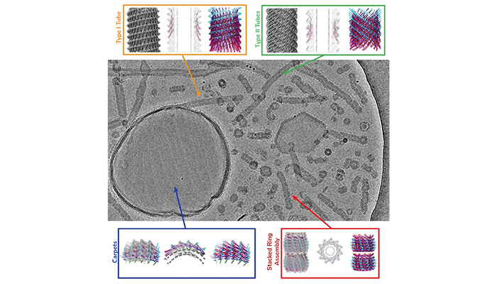
Cryo-EM shows a multitude of different Vipp1 structures: carpet-like structures, ring assemblies and tubes.
Credit: Forschungszentrum Jülich / Bendikt Junglas, Carsten Sachse

New insights into the structural mechanisms through which Vipp1, a membrane protein, repairs and stabilizes cell membranes in photosynthetic organisms have been uncovered by researchers using cryo-electron microscopy (cryo-EM).

Cryo-EM allowed the team from the Ernst-Ruska Centre for Microscopy and Spectroscopy with Electrons, Germany, to capture Vipp1 interacting with membrane surfaces in multiple structural forms. These forms include flat, carpet-like structures that stabilize the membrane surface, as well as ring and tube-like assemblies that appear to remodel damaged or stressed membranes. This detailed imaging provides a clearer understanding of how Vipp1 protects thylakoid membranes, which are essential for photosynthesis in plants, algae, and cyanobacteria.

Vipp1’s ability to form tubes filled with membrane material suggests that these structures may help isolate and remove damaged membrane sections. Additionally, Vipp1’s ability to form ring structures could facilitate the joining of membrane segments, stabilizing the overall structure of the membrane during stress.

In addition to the structural images, the study emphasized the importance of the protein’s α0 helix, which anchors Vipp1 to the membrane. Experiments showed that when this helix was truncated, Vipp1 lost its ability to interact effectively with the membrane, further highlighting its role in membrane protection.

The findings could have broader implications beyond photosynthetic organisms; for example, understanding how proteins like Vipp1 maintain membrane integrity under stress could inspire new approaches in biotechnology and medicine, particularly in the development of biomaterials or treatments for diseases involving membrane damage.

"This research provides valuable insight into how cells maintain their membrane structures, a process that is critical for many biological functions," the researchers concluded.

Newsletters

Receive the latest analytical science news, personalities, education, and career development – weekly to your inbox.

Newsletter Signup Image

False

Advertisement

Recommended

False

Related Content

The Analytical Scientist Innovation Awards 2024: #3
Microscopy
The Analytical Scientist Innovation Awards 2024: #3

December 6, 2024

4 min read

Bruker’s multiphoton microscopy module, OptoVolt, ranks third in our Innovation Awards. Here, Jimmy Fong, product development lead, walks us through the major moments during development.

Let Me See That Brain
Microscopy
Let Me See That Brain

December 9, 2024

1 min read

TRISCO sets a new standard for 3D RNA imaging, delivering high-resolution and uniform images to offer insights into brain function and anatomy

Loading the Molecular Dice
Microscopy
Loading the Molecular Dice

December 12, 2024

2 min read

How a framework for controlling molecular reactions at the atomic scale has potential implications for nanotechnology, pharmaceutical synthesis, and clean energy research

Found in Translation
Microscopy
Found in Translation

December 16, 2024

1 min read

Cryo-EM addresses a key question in gene expression: how ribosomes initiate translation

False

The Analytical Scientist
Subscribe

About

  • About Us
  • Work at Conexiant Europe
  • Terms and Conditions
  • Privacy Policy
  • Advertise With Us
  • Contact Us

Copyright © 2025 Texere Publishing Limited (trading as Conexiant), with registered number 08113419 whose registered office is at Booths No. 1, Booths Park, Chelford Road, Knutsford, England, WA16 8GS.