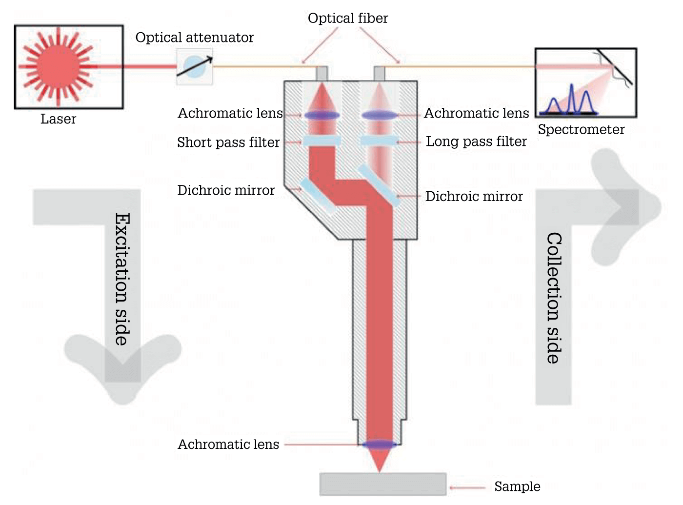
Oral squamous cell carcinoma (OSCC): common, but difficult to detect early – leading to poor prognosis. To address this challenge, researchers from the University Medical Center Hamburg-Eppendorf used shifted-excitation Raman difference spectroscopy (SERDS) to evaluate the molecular composition of OSCC, non-malignant lesions, and physiological mucosa – and whether they can be differentiated by Raman spectroscopy (1).
Based on physiological tissue of the oral cavity, they found that non-malignant and cancerous lesions can be distinguished with high accuracy using SERDS. The method also yielded high accuracy in identifying non-malignant lesions that required confirmation by surgical biopsy.
“Our study shows the potential of Raman spectroscopy for revealing whether a lesion is cancerous in real time,” said lead author Levi Matthies (2). “Although it won’t replace biopsies any time soon, the technique could help reduce the lapse of valuable time as well as the number of invasive procedures.”

References
- L Matthies et al., Biomed Opt Express, 12, 836 (2021).
- The Optical Society (2021). Available at: http://bit.ly/3tUVeXC.


