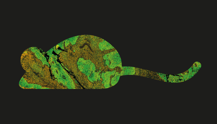
Glioblastoma accounts for over 60 percent of brain cancers, but survival five years beyond diagnosis is still rare. Raman and reflectance spectroscopies may allow us to distinguish healthy from diseased brain tissues with relative ease, improving our ability to detect and resect these tumors – and save patients.
After implanting murine glioma cells expressing enhanced green fluorescent protein into mice, Enrico Baria and colleagues used fluorescence microscopy to spot malignant tissue. Raman and reflectance spectroscopies aimed at the area of peak fluorescence intensity then informed a tissue classification algorithm, facilitating 97 percent identification accuracy when combining the two techniques.
The approach could provide a powerful tool for further animal studies – and could eventually complement preoperative MRI in humans.
References
- E Baria et al., Neurophotonics, 7, 045010 (2020). DOI: 10.1117/1.NPh.7.4.045010.


