Conexiant
Login
  • The Analytical Scientist
  • The Cannabis Scientist
  • The Medicine Maker
  • The Ophthalmologist
  • The Pathologist
  • The Traditional Scientist
The Analytical Scientist
  • Explore

    Explore

    • Latest
    • News & Research
    • Trends & Challenges
    • Keynote Interviews
    • Opinion & Personal Narratives
    • Product Profiles
    • App Notes

    Featured Topics

    • Mass Spectrometry
    • Chromatography
    • Spectroscopy

    Issues

    • Latest Issue
    • Archive
  • Topics

    Techniques & Tools

    • Mass Spectrometry
    • Chromatography
    • Spectroscopy
    • Microscopy
    • Sensors
    • Data and AI

    • View All Topics

    Applications & Fields

    • Clinical
    • Environmental
    • Food, Beverage & Agriculture
    • Pharma and Biopharma
    • Omics
    • Forensics
  • People & Profiles

    People & Profiles

    • Power List
    • Voices in the Community
    • Sitting Down With
    • Authors & Contributors
  • Business & Education

    Business & Education

    • Innovation
    • Business & Entrepreneurship
    • Career Pathways
  • Events
    • Live Events
    • Webinars
  • Multimedia
    • Video
Subscribe
Subscribe

False

The Analytical Scientist / Issues / 2021 / Jan / 3D Imaging of the SARS-CoV-2 Replication Cycle
Microscopy

3D Imaging of the SARS-CoV-2 Replication Cycle

01/13/2021 1 min read

Share

As the COVID-19 pandemic continues, so too do global efforts to learn more about the virus. Researchers at Heidelberg University investigated how SARS-CoV-2 interacts with its host, replicates within the cell, and leads to cell death (1). They used focused ion beam scanning electron microscopy to image infected cells at the subcellular level in 3D – identifying structural changes caused by the virus.

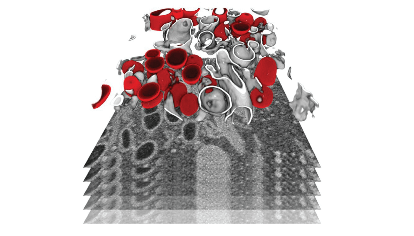
They found significant remodeling of the endomembrane system of infected cells – a structure that defines different compartments within the cell. This change enables the virus to produce its own replication organelles, with the viral genome then replicating in large amounts in double-membrane vesicles (shown in red above).

Credit: Julian Hennies/EMBL. Segmented subvolume of a cell, showing membrane-bound organelles (grey) and double-membrane vesicles (red).

Newsletters

Receive the latest analytical science news, personalities, education, and career development – weekly to your inbox.

Newsletter Signup Image

References

  1. M Cortese et al., Cell Host Microbe, Online ahead of print (2020). PMID: 33245857.

False

Advertisement

Recommended

False

False

The Analytical Scientist
Subscribe

About

  • About Us
  • Work at Conexiant Europe
  • Terms and Conditions
  • Privacy Policy
  • Advertise With Us
  • Contact Us

Copyright © 2025 Texere Publishing Limited (trading as Conexiant), with registered number 08113419 whose registered office is at Booths No. 1, Booths Park, Chelford Road, Knutsford, England, WA16 8GS.