A new microscope combines cryogenic transmission electron microscopy (cryoTEM) and liquid phase electron microscopy (LP-EM) within graphene liquid cells (GLCs) to achieve live imaging of biological processes in such detail that moving protein complexes are visible.
Imaging a dynamic biological process has proven difficult, due to the electron beam sensitivity of biological materials, their low electron contrast, and the necessity to repeatedly expose the system to the electron beam to monitor evolution in the process of interest.
“If you want to see protein complexes in such fine detail, you need an electron microscope,” explains Nico Sommerdijk, Professor of Bone Biochemistry at Radboud University Medical Center, The Netherlands, in a press release. “But the electron beam used can damage the biological material and the surrounding fluid, which is undesirable when you want to observe natural processes in the material over extended periods.”
The solution is to apply a protective layer around the material to minimize damage from the electron beam. This can be achieved with graphene. “But as soon as you apply it, the biological process you want to capture starts immediately,” Sommerdijk explains. “And then you have to quickly reach the microscope, locate the right spot in the tissue, and set up the microscope. This process takes at least half an hour, and sometimes the process is already over by then.”
The new technology, cryogenic-to-liquid phase correlative light electron microscopy (CL-CLEM), solves this problem with a multi-step workflow. First, the team apply a layer of graphene around the tissue and freeze it immediately, pausing biological processes. Then, using a light microscope, they identify the specific area in the tissue they want to visualize. Only after determining the correct orientation is the material placed in the electron microscope, which can perform measurements in liquid. In this setup, the material is warmed, reactivating the biological processes, which are then directly visualized on a nanoscale.
The researchers demonstrated this workflow's potential through the visualization of calciprotein particle (CPP) formation, a process relevant to vascular calcification. The technique enabled the researchers to capture early aggregation stages and monitor the development of CPPs over several minutes with nanometer resolution.
The technology sets the stage for future applications in real-time, in situ observation of delicate biological materials and processes in liquid environments.
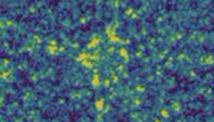
Credit: Nico Sommerdijk
Newsletters
Receive the latest analytical science news, personalities, education, and career development – weekly to your inbox.


