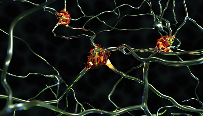
Suspicious protein molecules and fibrils aggregate on nerve cells in dementia.
Credit: Empa
Scientists have used advanced atomic force microscopy (AFM) to capture, for the first time, real-time, nanoscale images of amyloid-beta (Aβ-42) protein aggregates on fibrils, marking a significant step toward understanding the early mechanisms of Alzheimer’s disease progression. The study, led by Empa researcher Peter Nirmalraj and collaborators from the University of Limerick, tracked the formation of secondary nucleation sites on amyloid fibrils, where new aggregates form and potentially lead to cellular toxicity.
Aβ-42 proteins are known to misfold and clump into fibrils, forming dense structures that accumulate in the brain tissue of Alzheimer’s patients. Certain fibrils, termed “superspreaders,” were found to act as highly active nucleation sites, catalyzing further aggregation through surface features that bind new protein molecules. These superspreaders display rough, high-energy surfaces that facilitate continuous protein assembly, accelerating the formation of second-generation fibrils that may contribute to disease progression.
AFM, a high-resolution imaging technique, enabled the team to visualize these nanoscale interactions without the need for conventional staining. The researchers captured images of the fibrils in a salt solution, a medium that simulates the natural conditions of the human body. "This approach allowed us to observe the proteins in a state much closer to their native form," Nirmalraj noted in a press release. AFM provided important insights into the complex structure and activity of Aβ-42 fibrils, tracking their development over 250 hours and revealing how catalytic hotspots drive ongoing aggregation.
To validate the observations, the team paired AFM data with molecular model calculations, which supported the idea that specific surface textures enhance nucleation and the growth of aggregates. By classifying fibrils into subpopulations based on these surface characteristics, the researchers could distinguish between less active fibrils and those with a heightened potential for propagation.
The study helps reveal more about the underpinnings of Alzheimer’s pathology, highlighting the early aggregation sites as potential diagnostic and therapeutic targets.
Newsletters
Receive the latest analytical science news, personalities, education, and career development – weekly to your inbox.


