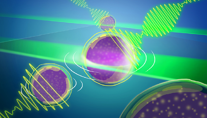
An artist's representation of the new Brillouin microscopy approach that allows entire light-sheets to interact with 3D biological samples. The scattered light reveals a unique optical interference signal that can be recorded with a custom-developed spectrometer, tremendously speeding up image acquisition.
Credit: Daniela Velasco/EMBL
A new microscopy technique that dramatically accelerates Brillouin imaging could enhance the study of biological materials while minimizing sample exposure to light. Researchers at the European Molecular Biology Laboratory (EMBL) have developed a full-field Brillouin microscopy system that offers a 1,000-fold improvement in speed and throughput, enabling rapid three-dimensional imaging of mechanical properties in living cells and tissues.
Brillouin microscopy is a label-free optical technique used to measure viscoelastic properties of biological materials. It relies on Brillouin scattering, a process where light interacts with natural thermal vibrations within a sample, causing a slight shift in frequency. This shift provides insight into mechanical characteristics such as elasticity and viscosity. However, conventional Brillouin microscopy techniques are hindered by weak scattering signals, requiring long acquisition times and high-intensity illumination that can damage biological samples.
To overcome these limitations, the EMBL team developed a new system that employs an imaging Fourier-transform spectrometer – Fourier-transform Brillouin Microscopy (FTBM) – that allows simultaneous spectral acquisition across an entire two-dimensional plane rather than scanning individual points. This improvement boosts throughput to approximately 40,000 spectra per second while maintaining a precision of ~70 MHz.
"We were on a quest to speed up image acquisition," said Carlo Bevilacqua, lead author of the study and an optical engineer at EMBL, in a press release. "Over the years, we have progressed from being able to see just a pixel at a time to a line of 100 pixels, to now a full plane that offers a view of approximately 10,000 pixels."
The technique provides a practical solution for imaging biological systems that are sensitive to prolonged exposure to light. By distributing laser illumination across a broad field of view, FTBM reduces the phototoxic effects seen in traditional confocal Brillouin microscopy methods. The researchers demonstrated its effectiveness by imaging both synthetic material phantoms and living zebrafish larvae, highlighting the technology's potential for studying biomechanical properties in vivo.
According to Robert Prevedel, senior author of the study, the development is akin to past revolutions in microscopy: “Just as the development of light-sheet microscopy here at EMBL marked a revolution in light microscopy because it allowed for faster, high-resolution, and minimally phototoxic imaging of biological samples, so too does this advance in the area of mechanical or Brillouin imaging.”
The researchers plan to further optimize the technique by integrating nanostructured surfaces and metamaterials to improve imaging stability and enhance contrast. "We hope this new technology – with minimal light intensity – opens one more 'window' for life scientists' exploration," Prevedel added.
Newsletters
Receive the latest analytical science news, personalities, education, and career development – weekly to your inbox.


