Newsletters
Receive the latest analytical science news, personalities, education, and career development – weekly to your inbox.

False
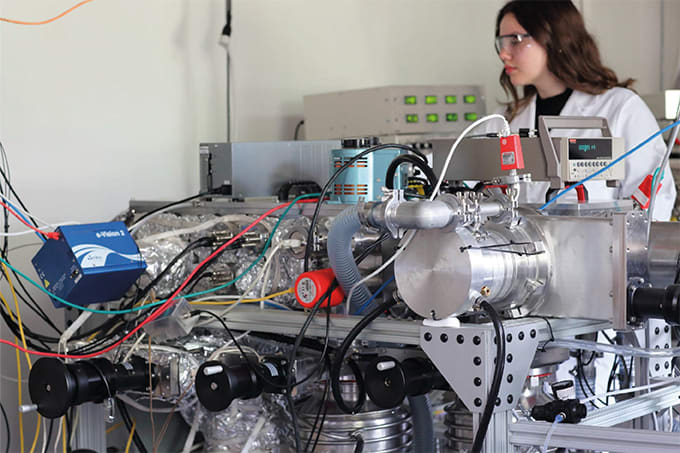
Spectroscopic analysis reveals lipid–RNA interactions governing nanoparticle formulation efficiency
Receive the latest analytical science news, personalities, education, and career development – weekly to your inbox.

December 12, 2024
2 min read
How a framework for controlling molecular reactions at the atomic scale has potential implications for nanotechnology, pharmaceutical synthesis, and clean energy research
October 1, 2024
10 min read
Charge-detection mass spectrometry (CD-MS) has extended the range of MS to gigadalton-sized viruses and polymers; and with a commercial instrument in development and exciting new applications in complex protein mixtures, maturity beckons

October 1, 2024
1 min read
Researchers develop an NMR method to distinguish between enantiomers without the need for chiral agents

October 2, 2024
2 min read
Researchers combine tissue imaging with proteomics to shed light on the neurotoxic effects associated with HIV medication Efavirenz treatment
False