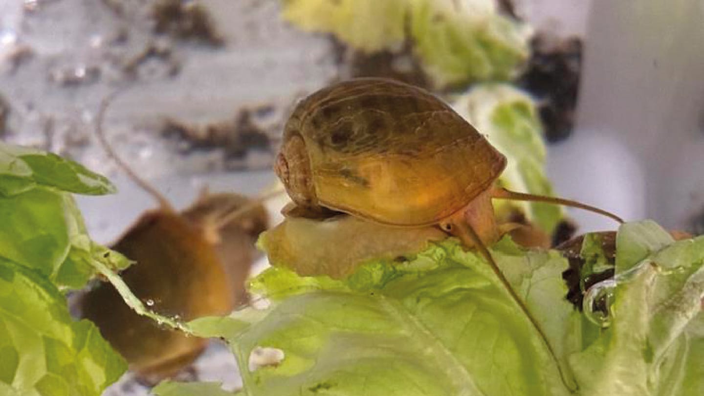
The capacity to regenerate damaged tissues varies widely across the animal kingdom, but the ability to reconstruct an entire, fully functional eye has remained undocumented in complex organisms. Now, researchers from the Stowers Institute for Medical Research, Kansas City, USA, have demonstrated that the apple snail (Pomacea canaliculata) can regenerate its adult camera-type eye following complete removal.
Using CRISPR/Cas9 tools, transcriptomic profiling, and microscopy, the team tracked the regeneration process from early wound healing through to the restoration of photoreceptors, retinal layering, and neural reconnection. Their analyses revealed that many of the same gene networks active during embryonic eye development are redeployed during regeneration – but in distinct, non-linear patterns. The work establishes P. canaliculata as a genetically tractable model for investigating complex organ regeneration and provides a molecular framework that could guide future studies of sensory repair.
To learn more about the inspiration behind the research, the analytical challenges involved, and the broader implications for regenerative biology, we spoke with Alejandro Sánchez Alvarado, senior author of the study.
What inspired you to study eye regeneration in snails?
Eyes are among evolution’s greatest achievements: intricate masterpieces that have evolved in astonishing forms across the animal kingdom. In regenerative biology, restoring sensory organs such as the eye remains one of the most challenging frontiers. While several organisms can regenerate parts of the eye, like the lens in salamanders or the retina in fish, no model existed to study the regeneration of an entire, fully functional adult eye.
Our work with Pomacea canaliculata revealed something extraordinary: this species, equipped with a sophisticated camera-type eye, can completely regenerate the organ – an ability previously thought limited to creatures with far simpler visual systems. Coupling this with CRISPR/Cas9 technology has given us, for the first time, a genetically tractable system outside the usual vertebrate and insect models to explore how such a complex organ is assembled, regenerated, and reintegrated into the nervous system.
What was the biggest hurdle you faced and how did you overcome it?
The biggest challenge was managing and making sense of the immense volume of data from our gene expression analyses. We collected samples at multiple time points, each generating millions of data points, and then compared these across the entire regeneration timeline. This required us to systematically identify which genes were turned on or off at different stages. Through sophisticated bioinformatic analyses, we were able to pinpoint candidate genes likely responsible for orchestrating eye regeneration, turning an overwhelming dataset into a clear roadmap of molecular events.
Did anything surprise you during the research – perhaps something that contradicted assumptions or revealed something unexpected? Any “eureka” moments?
Absolutely. The breakthrough was not just in observing an eye regrow from its deepest photoreceptors to the neural connections with the brain, but in uncovering the genetic choreography behind it. We found that the same core gene networks used during embryonic eye development were redeployed during regeneration, yet in non-linear, asynchronous ways. This revealed a striking flexibility in how evolution reuses its genetic “playbook,” modifying ancient programs to meet the challenge of rebuilding a complex sensory organ from scratch.
The study integrates genomic analysis, microscopy, and developmental biology. What role did interdisciplinary collaboration play in bringing all these tools together?
Inter- and transdisciplinary collaboration were absolutely essential. Regenerating a complex organ like the eye involves changes that are both molecular and structural, unfolding across time and space. We needed quantitative tools to measure gene expression and qualitative tools to visualize those changes at the tissue level. Integrating these distinct datasets required reducing their dimensionality in ways that allowed direct comparison, enabling us to generate new models and hypotheses. This synthesis was only possible through the combined expertise of genomic scientists, developmental biologists, and imaging specialists working toward a shared goal.
What do your findings suggest about the possibility of regenerating complex sensory organs in humans?
Our findings demonstrate that it is biologically possible to regenerate a complete, fully functional adult camera-type eye. Understanding the full molecular and cellular blueprint for this process could open new avenues for treating eye injuries and degenerative conditions in humans. While translating this knowledge into medical reality will require significant further research, we now know that the blueprint exists, and that we can begin the work of decoding and adapting it.
What are your future plans for this work?
Our next goal is to map the regeneration process with greater precision by effectively decoding the “instruction manual” Pomacea canaliculata uses to rebuild its eyes. By identifying the fundamental biological principles underlying this ability, we hope to apply them to species (including humans) that currently lack such regenerative capacity, ultimately bringing the repair and restoration of complex sensory organs within reach.
Newsletters
Receive the latest analytical science news, personalities, education, and career development – weekly to your inbox.