Splitting Hairs
Despite the recent bad press, cutting-edge analytical techniques are transforming the field of hair analysis
Bryn Flinders |
Forensic scientists adopted microscopic hair analysis way back in the 1950s, examining the characteristics of forensic hair samples in terms of pigment distribution or the scale pattern in order to compare it with hair taken from a suspect.
However, the validity of the technique came under heavy fire earlier this year. An article in The Washington Post reported that US Federal Bureau of Investigation (FBI) examiners gave flawed testimony regarding microscopic hair analysis, resulting in several wrongful convictions (1). But in a way, that’s old news. The introduction of DNA profiling completely changed the game, and convictions based on microscopic hair analysis have been overturned as a result.
Moreover, hair analysis continues to evolve and it can provide a great deal of useful information. For example, toxicologists and forensic scientists use it to detect drugs of abuse. Hair analysis is highly advantageous as it can provide a historic record of ingested chemicals versus a transient detection in biological fluids, and it can reveal chronological information about drug intake. This approach uses chromatographic techniques coupled with mass spectrometry to analyze 1-cm long hair segments. As good as this technique is, sample preparation is complex and also consumes the sample, making repeat analyses difficult.
Recently, the use of mass spectrometry imaging (MSI)-based techniques, such as matrix-assisted laser desorption/ionization (MALDI) and secondary ion mass spectrometry (SIMS), have been used to provide visual, chronological information on the distribution of pharmaceutical compounds or drugs of abuse in hair samples (see Figure 1). The improved spatial resolution results in a more accurate drug usage history, using micrometer versus centimeter lengths of hair, and in hours versus months.

Figure 1. MALDI-MS image showing the distribution of cocaine throughout five hairs.
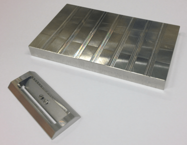
One of the issues often raised is whether detected compounds are present because of external contamination or actual abuse. The use of decontamination protocols and the detection of unique metabolites is usually enough to discern this. An innovative apparatus (developed by FOM-AMOLF/M4I, see Figure 2) to prepare longitudinal sections coupled with high spatial resolution imaging techniques provides more detail for the consequences of decontamination protocols (2).

Figure 2. Innovative apparatus (cutter and block) used to literally split hairs.
Mass spectrometry imaging has potential in the future to provide information about a suspect from single hair samples found at a crime scene, because the presence of pharmaceuticals, drugs of abuse, or other compounds could be unique to a suspect. If a DNA profile is unobtainable due to the absence of the hair root, or links need to be made to a particular chemical exposure, the technique offers real advantages. Chemical analysis of hair samples could potentially provide valuable information on proteins, metabolites or small molecules, for example, that together with other evidence could help to identify a suspect.
- goo.gl/UMn9cI
- B Flinders, et al., “Preparation of longitudinal sections of hair samples for the analysis of cocaine by MALDI-MS/MS and TOF-SIMS imaging”, Drug Testing and Analysis, first published online (2015).
After receiving his BSc in Forensic and Analytical Science in 2009 from Sheffield Hallam University, UK, Bryn Flinders began his PhD project on the application of MALDI-MSI to monitor the distribution of respiratory compounds in the lungs at the same university under the supervision of professor Malcolm Clench. Then in 2013, he took up a postdoctoral position at FOM Institute AMOLF in Amsterdam, the Netherlands under the supervision of professor Ron Heeren. His area of research was the application of mass spectrometry imaging to monitor the distribution of drugs of abuse in hair. Recently, he moved to the Maastricht Multimodal Molecular Imaging Institute (M4I) where he continues his research.


