This Week’s Spectroscopy News
Georg Ramer’s extra special EV method, fresh perspectives on how we perceive and manage pain, and a new method for predicting beef aging time…
James Strachan | | 3 min read | News
Essential Reading
Extra Special EV Protocol
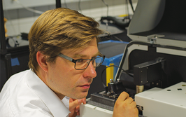
One paper that stood out this week: Georg Ramer and colleagues at TU Wien unveiled a new method for analyzing individual extracellular vesicles (EVs). EVs are tough to study because of their small size, heterogeneity, and the limitations of bulk or label-based techniques.
The team addressed these challenges using atomic force microscopy infrared spectroscopy (AFM-IR), combined with antibody-based capture, hyperspectral imaging, and machine learning. Their workflow allows label-free chemical profiling of dozens of EVs at once, revealing striking sub-vesicle heterogeneity and setting the stage for more robust, high-throughput EV analysis.
As it happens, we spoke with Ramer about the trends and challenges for the AFM-IR field back in 2013. This is what he had to say:
“Every year, we see new measurement modes coming out and ever smaller samples (single molecule!) are being analyzed. However, if this technique is to be even more widespread, it needs to become a tool for routine analysis that can be operated in industrial quality control labs and by researchers from other fields. In those applications, having record-breaking sensitivity or one less nanometer of spatial resolution is not required. Rather, the results must be reliable and reproducible.”
Also in the news…
Researchers develop a scatter-free UV/visible spectroscopy method that enables rapid, accurate quantification of RNA in lipid nanoparticles, overcoming the light scattering issues that hinder traditional UV/vis techniques and outperforming commonly used alternatives such as RiboGreen in both precision and simplicity. Link
Near-infrared spectroscopy, combined with chemometric modeling, accurately predicts beef aging time, offering a rapid, nondestructive alternative to conventional meat quality assessment methods. Link
Smart surface-enhanced Raman spectroscopy, integrated with machine learning, reveals how nanoplastics and their “carrier effects” disrupt cellular metabolism and extend cell cycle phases, offering a dynamic, in situ approach to studying nanoparticle toxicity in real time. Link
Visible–near-infrared spectroscopy, combined with machine learning, accurately classifies cotton Minicard stickiness into four levels, offering a rapid, nondestructive alternative to traditional stickiness tests. Link
The Analytical Scientist Presents:
If you're enjoying this article, you may like to join our mailing list to receive new content every fortnight. The Spectroscopy Newsletter offers the hottest topics at your fingertips, specially chosen by our wonderful Editorial team!
The Spectacular and Strange
Pain on the Brain
Two intriguing studies offer fresh perspectives on how we perceive and manage pain – especially in patients who can’t easily communicate their discomfort. In one study, researchers explored how the brains of patients with disorders of consciousness respond to pressure-induced pain. Although traditional signs of brain activation were minimal, functional near-infrared spectroscopy (fNIRS) revealed increases in functional connectivity between sensory, motor, and cognitive areas during stimulation – suggesting that even in the absence of overt responses, the brain might still be coordinating a response to pain
Meanwhile, a team working with cancer patients used fNIRS in a different context – to classify pain severity and evaluate the soothing effects of a virtual reality ocean journey. Their machine learning model could accurately classify pain into three levels based on brain signals, and the immersive VR experience led to significant reductions in perceived pain for most participants. Functional connectivity in the brain also shifted after VR, hinting at real neurophysiological changes behind the relief.
Also from The Analytical Scientist
Microplastics Detected in Cat Placentas and Fetuses
Microplastics have been found in the placentas and fetuses of stray cats during early pregnancy. Of the eight cats studied, microplastics were detected in placental tissue from three cats and in fetal tissue from two. Raman spectroscopy allowed identification of both the polymeric backbone in some cases and, more frequently, the associated pigments.
“Micro-Raman spectroscopy is one of the most used techniques for detecting and identifying micro- and nanoplastics in environmental and biological samples,” the authors note, adding that while the technique can underestimate total particle count – especially for colourless plastics – its specificity in identifying both polymers and pigments makes it a valuable tool for tissue-based studies. Read more!

Over the course of my Biomedical Sciences degree it dawned on me that my goal of becoming a scientist didn’t quite mesh with my lack of affinity for lab work. Thinking on my decision to pursue biology rather than English at age 15 – despite an aptitude for the latter – I realized that science writing was a way to combine what I loved with what I was good at.
From there I set out to gather as much freelancing experience as I could, spending 2 years developing scientific content for International Innovation, before completing an MSc in Science Communication. After gaining invaluable experience in supporting the communications efforts of CERN and IN-PART, I joined Texere – where I am focused on producing consistently engaging, cutting-edge and innovative content for our specialist audiences around the world.
