I started my career in capillary zone electrophoresis (CZE) instrument development with laser-induced fluorescence detection, for environmental applications. While attending the Microscale Separations and Bioanalysis Conference in Canada in 2016 to present my research, I went to a forensics session and was shocked to hear about the major backlog in sexual assault cases in the US. As it stands, there’s tens of thousands of rape kits awaiting analysis in the US – that’s tens of thousands of victims whose lives could be on hold while they await justice. The injustice of it deeply affected me. Afterwards, I asked the presenter if anyone had attempted the separation using capillary electrophoresis. He said, “No! Why don’t you give it a try?” So I started working on it in my spare time.
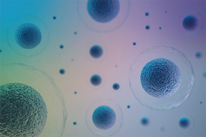
Scientifically, this is a separations issue; the main challenge in the analysis of sexual assault kits is the separation of sperm cells (containing male DNA), from the epithelial cells in the sample. The epithelial cells grossly outnumber the sperm cells in most samples, which makes it difficult to get a clean DNA profile of the perpetrator. The current method of differential extraction is very inefficient. The process uses a series of solutions and detergents to wash the sample off the swab, targeting the fragile epithelial cells first and the hardier sperm cells last. However, each of these fractions contain DNA from both perpetrator and victim and produce mixed profiles that are often difficult to interpret. Furthermore, the process requires a lot of analyst interaction with the sample, which increases the risk of sample contamination or loss. CE is already used in every crime lab for DNA analysis, and is known to produce efficient separations of DNA and small molecules. In my previous work, I pushed the upper limits of CZE by separating mixtures of bacteria for environmental applications. But could CZE be used to separate mixtures containing epithelial cells, which were over 40 times the size of the E. Coli I was previously working with? The scientists I spoke with at the conference were concerned that the epithelial cells would clog the capillary. In response, I spent a few months working on sample preparation – testing different buffers to remove the sample from the swab, and manipulating CZE injection and separation parameters to overcome this challenge. Then, I interfaced the CZE separation with an automated fraction collector developed in my lab. I could then inject mock sexual assault samples into the CZE system, separate intact sperm cells from epithelial cells and lysed cellular debris, and collect purified fractions. With this technology, we can get very specific separation in under 15 minutes, and I’m continually striving to achieve an even faster separation with equal efficiency. This is a more effective alternative to the current method of differential extraction, which requires samples to incubate for a few hours and often overnight. Furthermore, there is very little human interaction with the sample since there are no wash steps. I’ve been using a visual analysis method to quantitatively determine my yield and evaluate the separation, but I would like to switch to something more in-depth such as real-time PCR coupled with fluorescence detection in the near future.
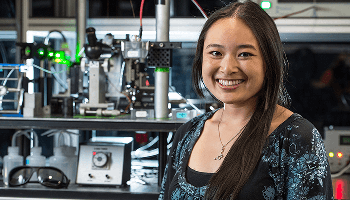
The University of Notre Dame’s Tech Transfer Office will be looking to commercialize the technology. My job is to improve the instrument and to continue running experiments to show that it not only can work on fresh samples, but it can also handle the backlog. I’m currently doing a time study to ensure system effectiveness with three-month-old mock sexual assault swabs – I’d like to go back up to a year and test different storage conditions (temperature and humidity) since many counties do not have ideal storage facilities. I aim to demonstrate that speed, simplicity and sensitivity make this method worthwhile for every lab. The University of Notre Dame does not have a forensics program, so everything I’ve done has been very reliant on collaboration and communication with other research institutions. The forensics community have been very supportive. We’re all passionate about finding ways we can help people – that’s what we’re in this job for. It’s not about fame, making money, or beating your competition – it’s about working together to solve society’s problems. I’m very hopeful about the future. There are a lot of people passionate about making progress in forensic science, bringing justice to our communities and lowering crime rates – I want to be part of that. Sarah Lum is Bioanalytical Chemistry PhD student and Graduate Research Assistant at the University of Notre Dame, Indianapolis, USA.

I became enamored with acoustic differential extraction (ADE) at graduate school at the University of Virginia. I joined the Landers Research Lab in 2014, and I have since been working on the development of a microfluidic technology (SONIC) that uses acoustic force to separate sperm cells from epithelial cells in sexual assault samples. Small-scale chemistry The SONIC system originates from a collaboration that started with Prof Thomas Laurell at Lund Univ and incorporates ADE on a microfluidic device – essentially using sound waves to apply pressure and separate particles. The acoustic trapping principle is the application of a standing sound wave through a microfluidic channel filled with liquid. Those sound waves create low-pressure nodes where they intersect, and high-pressure anti-nodes where the sound waves are out of phase. If you flow particles through that acoustic trapping site, they’ll follow the path of least resistance into the low-pressure nodes. And if you tune the frequency of the sound waves properly, you can actually trap and hold particles of a certain size, while everything else flows around it. Different cell types in the human body vary drastically in terms of size, shape and function. Sperm cells are very well conserved across humans – they’re all around 6.0 micrometers in size (at the head) and ~50 micrometers long (head-to-tail), , with roughly the same shape and features. That means we can tune our trapping site very precisely to sperm cells. Once we’ve flowed our sample through and are holding those sperm cells in place, we have multiple downstream avenues that go to different chambers; we can let all of our sample waste go to one, then switch the flow and release sperm cells into another, thereby purifying those cells that we want to capture. The conventional method used to separate sperm cells from other cells (primarily epithelial cells) is simple differential extraction. You spin your sample containing multiple cell types at 18,500 x g for 10 or 12 minutes, and the sperm cells will pellet out to the bottom. The analyst removes the supernatant, re-suspends it, and repeats this spin and wash step until they get a purified sperm fraction. It still surprises me that conventional analysis is so manual and thus, how variable this can make the process in handling these types of samples. In essence, what we’re trying to do in the Landers Lab is automate that separation process – taking it out of the hands of the user to make it more uniform. With our methods, you simply load your sample; the metering, fluidic control, trapping, and manipulation are all handled by the instrument – and you are presented with a small vial of purified sperm cells from your sample. Baby steps The response to SONIC from the community has generally been positive, although people don’t always appreciate the steps that need to be taken in a project like this. When I describe it to other forensic or analytical scientists, they often jump straight to posing convoluted scenarios: “What if you get a sample that has cells from five different people, with four different suspected attackers?” I have to explain that we’re not addressing that yet; it takes baby steps to get to that point. What we’re doing might not change the types of samples you can look at, but it could open the door to more reproducible male capture – and, in this field in particular, that’s crucial. One of our biggest challenges – and this was unexpected – has been getting reliable information from the rest of the forensic community. We don’t have access to real casework. It was really hard, for example, to find out the ratio of female to male cells in a typical sample – we were given numbers that ranged from 1:1 to 600:1.
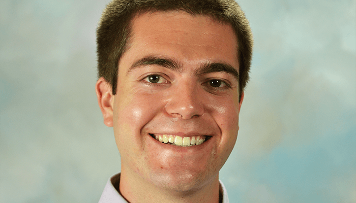
Probing on Palm Beach An exciting new development for me was going down to work with Palm Beach County Sheriff’s Office (PBSO) in Florida, to observe some of their forensic techniques, train them on using the instrument that we developed, and then compare different extraction methods. They handed me a list of adjudicated samples – tank tops, sheets, condoms, cheek swabs – all kinds of samples and substrates and cell types that I wasn’t ready for. It was much more of a challenge than I thought, but a great opportunity to try the instrument with real samples. One gratifying moment was when they presented us with an adjudicated sample – a cutting from a sheet that had been stored since 2009. We pulled it off the shelf, resuspended it, and were able to separate sperm cells from that sample using the instrument. From our sample, we were able to generate a DNA profile that matched the reference profile that they obtained via their own method eight years earlier. Perhaps not the most challenging sample, but a great moment for us nonetheless. The trip was really eye-opening. It struck me how unique every lab is; there are different national and state guidelines on how you handle samples, and how you handle these types of investigations. PBSO is a very well-funded state lab, so they have the best instrumentation. It seems like other labs who have obtained less funding may not be able to handle as many samples or hire as many analysts – which means that having new technology that expedites analysis is even more important. Translating forensics I’d really had no exposure to forensics before working with this group, but what really hooked me was how easy it is to convey the importance of what I’m working on. Everyone I talk to agrees that it’s important to help address the backlog of samples in solving these crimes by speeding up the analysis process. Forensics is in some ways more visible than other areas of analytical science. Does our technique have scope beyond forensics? We believe so. A recently graduated student from our lab has applied this acoustic isolation technique to the separation of cancer cells. Circulating tumor cells appear in very low numbers in the bloodstream; if you can focus on the differences of those cells – be it in size, shape or compressibility – and separate them using our acoustic technique, then you have the potential to tailor the treatment to the type of cancer the patient has. It’s the same principle, but a whole other set of parameters and instrumentation being applied to a new field. Charlie Clark is a PhD candidate at the Landers Group, Department of Chemistry, University of Virginia, USA.
The Science and Nothing But the Science, with Craig O’Connor
One Piece of the Puzzle, with Kacey Cliburn
Fly in the Face of Evidence, with Jennifer Rosati
Battling the Backlog, with Sarah Lum and Charlie Clark


