Skeleton Crew
Raman spectroscopy identifies rickets in 400-year-old bones from the Mary Rose
The Mary Rose – a famous English warship – sank in 1545. Despite major excavation only beginning in 1979, the bones of the crew – and the chemical information held within – have been well preserved thanks to a covering of silt. Indeed, the chemical fingerprints from the bones from the Mary Rose were comparable to those obtained from fresh bone samples.
The research team, based at University College London, was originally working to develop Raman spectroscopy to study bone disease in living patients at the Royal National Orthopaedic Hospital when they were asked by Alex Hildred from the Mary Rose Trust whether the technique would work on archaeological specimens.
“The rickets work first came about after a discussion about modern-day rickets in London,” says Kevin Buckley, one of the authors of the study (1). “Since the Mary Rose study, we have begun to scan the bones of individuals suffering from rickets at the Royal National Orthopaedic Hospital. It is hoped that some of the chemical information that we obtained from the 16th-century sailors could have a bearing on our studies of rickets in children in the 21st-century.”
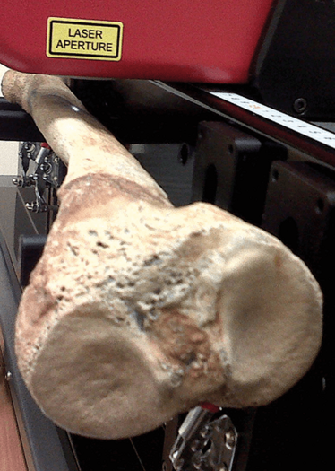
Raman data were collected from ten bone samples from the Mary Rose – five normal tibiae and five bowed tibia – and fresh comparison spectra were obtained from a cadaveric sample from Bristol University. “We used an 830 nm laser with ~300 mW power onto the sample to obtain the chemical fingerprints for the tibiae,” explains Jemma Kerns, lead author of the study. “The bones were held on a platform and we scanned along the length of the anterior aspect of the tibiae in 1 mm intervals. The bones that were suspected to be from individuals with metabolic bone disease (because of their warped shapes) had abnormal chemical compositions.”
The team isn’t finished with their work yet; they will investigate more bones from the Mary Rose that indicate the presence of other bone conditions; they have also been approached by other museums to discuss similar projects.
“The week before the Mary Rose Paper was accepted we also published a case study in BoneKEy, which showed that Raman spectroscopy, specifically Spatially Offset Raman Spectroscopy (SORS), could be used to detect bone disease in vivo (once a chemical abnormality was known to be present) (2). We are now working towards a larger study to detect bone disease in cohorts of individuals and hope to publish a progress report soon,” says Buckley.
- J. G. Kerns et al., “The Use of Laser Spectroscopy to Investigate Bone Disease in King Henry VIII's Sailors”, J. Archaeol. Sci. 53, 516-520 (2015).
- Kevin Buckley et al., “Measurement of Abnormal Bone Composition In Vivo Using Noninvasive Raman Spectroscopy”, IBMS BoneKEy 11 (2014). DOI: 10.1038/bonekey.2014.97
"Making great scientific magazines isn’t just about delivering knowledge and high quality content; it’s also about packaging these in the right words to ensure that someone is truly inspired by a topic. My passion is ensuring that our authors’ expertise is presented as a seamless and enjoyable reading experience, whether in print, in digital or on social media. I’ve spent seven years writing and editing features for scientific and manufacturing publications, and in making this content engaging and accessible without sacrificing its scientific integrity. There is nothing better than a magazine with great content that feels great to read."
